0:02
SNI and SNI Digital Baghdad Neurosurgery Online Meeting, held on October 23, 2022, the meeting on October 23, 2022, the meeting originator and coordinator with Samir Hawes, universities of
0:19
Baghdad and Cincinnati.
0:29
The meeting subject is neural oncology in Iraq. Part 1. The historical perspective, 40 minutes. Lecture and discussion, LD.
0:46
Part 2. Challenging cases from Iraq. Personal experience from two neurosurgeons, 60 minutes. LD. Part 3. Update on neural oncology worldwide. 60 minutes. LD. The speaker will discuss
1:09
highlights in history of neural oncology with emphasis on practice and research in Iraq.
1:19
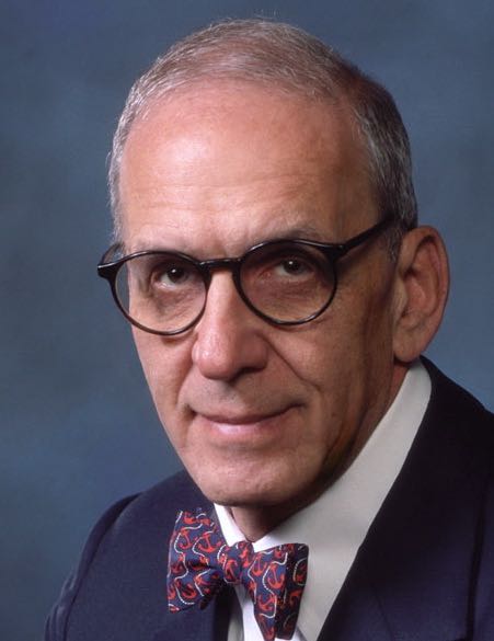
The speaker is Professor A. Hadi Akalili.
1:25
Former Chair of the Department of Neurosurgery at Baghdad University.
1:33
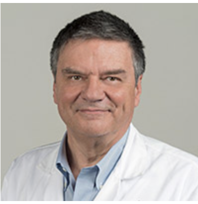
The speaker on the subject of neuro-oncology through interesting cases is Dr. Moneer K. Faraj, President of Arabic Board of Neurosurgery in Iraq, Department of Neurosurgery, College of Medicine,
1:54
University of Baghdad, Iraq.
1:58
The speaker will discuss neuro-oncology
2:11
through interesting cases, Dr. Warmidith E. Matai, Department of Neurosurgery, Neuroscience Hospital, Baghdad, Iraq
2:16
The speaker today is Dr. Terral Patel, Department of Neurosurgery, University of Texas Southwestern Medical Center, Dallas, Texas, United States of America. The target audience is students,
2:34
residents, young neurosurgeons, experienced neurosurgeons, and basic scientists
2:43
The video editors were Mustapas Ishmael, College of Medicine, University of Baghdad, and Fatima Ayad, fourth year medical student, University of Baghdad. Hi everybody, so you are welcome here.
3:03
Today we have the 18th Surgical Neurology International Baghdad Meeting This is like 18 months from starting of this event. It started initially as an collaboration between the United States and a
3:19
working team from Iraq, and now we are doing extra milestones with each meeting, try to make more collaboration, a wider area to coverage, and the most important to share experience from different
3:36
parts of the world So this is the first meeting that focus on a specific topic. And this is a new trial for us to focus on a specific speciality try to,
3:52
to give together what's there in Iraq from background, then what's happening now in Iraq regarding your oncology, and we have also a speaker. We have a guest speaker from the United States, just
4:09
also to give us a more update on the same topic. And so regarding the program, so the first talk will be from Dr Salini, and then Dr Romoneer, then Dr Romone on
4:24
the last talk will be from Dr Patil. And through the talks, we will try to have a five minutes question and answer after each talk. And then at the end of meeting, we can have extended questions
4:39
and answer opinion comments
4:42
according to their topics. So welcome everybody and I invite Dr Osman to start the introduction for the meeting. The meeting subject is neuro-oncology in Iraq. Part one, the historical perspective
5:01
40 minutes. lecture and discussion, LD. The speaker will discuss highlights in history of neuro-oncology with emphasis on practice and research in Iraq.
5:17
The speaker is Professor A. Hadi
5:21
Al-Khalili, former chair of Department of Neurosurgery Bagdad University For
5:31
Hadi, you're a towering national figure in Iraq and in the Middle East have tremendous respect for you and can you carry on from here? Yeah, thanks. Chair, a couple, thank you for the
5:46
encouragement for the introduction and for this great project, the noble project to helping people, the young view resurgence all over the world With your leadership, with your expertise and with
6:00
creating
6:03
international and the Baghdad chapter of it.
6:11
Mighto could be just highlights on history of neuro-oncology with emphasis on practice and research in Iran.
6:21
The further backward you can look, the further forward you can likely to see, and that's what is in Churchill. So studying history of the specialty or history of anything is useful really to open
6:37
eyes for better
6:41
productivity.
6:43
I will touch this on Mesopotamia medicine, Egyptian medicine, the Greek medicine, Muslim Arabian medicine, medieval, and modern times, and then come to Iraq practice, and then also I will touch
6:54
on the cultural difference of the informed content.
7:00
Cancer in Latin means crab, and onco, again in Latin means tumor, tumor means swelling andglaya meansglue andgreet Thanks.
7:15
In
7:21
Mesopotamia and
7:25
medicine, we have this wonderful book of Serlock and Anderson, about more than 800 pages of the book which is studying only the diagnoses in Babylonian and Syrian medicine. They don't touch on
7:38
treatment, just the diagnosis So in the tumor section, they said, If a falling spill upon him, and it has been falling upon him for one year, and at its sign, it comes over him, its worrisome,
7:53
if seizure comes closely spaced, and in the middle of the day, it will be difficult for him.
8:02
In another statement about praying tumor in that book, if he is sick with mild headache, And just as an ox continually looks at his. own feet and looks at his own feet, that patient will not do
8:18
well, indicating of gaze paralysis or something.
8:24
Edwin Smith in his papyrus about
8:29
Egyptian medicine, there
8:34
was no referral to brain tumors, only for cancer of the breasts in the Egyptian medicine. I could not see anything any referral to brain tumors. Hippocrates was the first one to call it carcinoma,
8:49
which is carcinoma, and it looked as it looked like crab, and then gallon, viewed cancer as Hippocrates had, and considered the patient is incurable after diagnosis of cancer.
9:03
In the Muslim Arabian medicine, we know that arises, these leading three figures in medicine, and of course there are a lot more. They are leading figures in medicine in the first millennium and
9:18
just after. And the most important one, for our case, is Al Bukhasis, who is Abu Ghassam Zarawi. And he was considered by one author in the states that the father operative surgery. And in this
9:35
picture, he's showing that he's operating on the head And he improvised more than 200 surgical instruments, including for cranial surgery or instruments.
9:50
But then later on, Amrah Paray, who is considered the father of modern surgery
10:03
in the 16th century, and then he developed these tools to properly open the head
10:06
Then we have Sir
10:09
William McEwen, McEwen, who is a wonderful person. person if one studies his life, and he was the first perform removal of Frontal Meningioma in 1879. And in
10:24
one of the lectures in Glasgow, Harvey Cushing was saying about
10:30
him that we merely stand on,
10:37
we merely stand on the shoulders of our predecessor, the Stradi contemporary figures of McEwen on one side, and horsely on the other side, supporting the arch of modern neurology surgery. So he
10:52
considers these guys as the pillars.
10:56
There are so many nice stories about McEwen, his achievement at his work.
11:04
And then we have Sir Rickman
11:07
Godley, who was the first to operate. on brain tumors in 1884 and diagnosed and operate on this tumor, but was reported in 1885 in this
11:23
article.
11:25
The three giants of modern neurosurgery are Victor Horsley and Harvey Pushing and Walter Dandy. Of course, there are so many other giants in the book of neurosurgical history of feet, of clay and
11:40
iron, which possibly have seen that book.
11:45
The important discovery is Mary Curie and her husband, Pierre Curie, discovered radium, and they got Nobel Prize for that in 1903. Their daughter, Irene, and her husband, they artificially
11:59
created radioactive atoms, and for that they got Nobel prize in 1935.
12:06
And I, myself, used this radium needles when I was a resident for a patient with cancer of the tongue.
12:18
Now, chemotherapy was started for cancer by Paul Eillich, and he got Nobel Prize in 1902,
12:26
while immunotherapy started by William Coley in 1890, and the radiotherapy was started by M. L. Corropa in 1996.
12:53
Breaky therapy in 1966, Mundinger implanted a radium 192 wires into low low grade glioma. And Gerhard Friedlander used IOD in 125 implanted in 1979 in
13:11
gliomas.
13:14
Practice in Iraq, in general, neurosurgery of service in Iraq started in 1946 She was trauma, but then it developed including tumor surgery and specialized hospital in 1969 and then 1990 and then
13:28
more centers and developed since then.
13:32
Our means of diagnosis at that time and our time when we practice at the beginning was plant, plane, radiography, and pneumonia, encephalography and then cerebral angiography with direct puncture
13:43
of the carotid artery and the the technetium brain scan
13:48
and then Let's see if this can came at 90.
13:53
in 1978, you can see this is the vague picture of the skin, which we was to depend on for surgery on tumors.
14:03
Treatment in general, we use to use, of course, biopsy only maybe or surgical resection partial or total, if possible. And the radiation therapy, we only had cobalt, 60. And we put marks on
14:15
the head, so the direction of the radiation would be on that mark all the time when you come to different sessions. Then we have the accelerator with cobalt, but because of the sanction, cobalt
14:30
was prevented from arriving to Iraq. So it was only the accelerator. And we did not have any chemo therapy there.
14:40
Surgery was classical, nothing fancy about surgery We just used that, and blew up a very simple means of doing that. surgery. But then in the 90s, we have the Kuza aspirator, and we have the CO2
14:56
laser. And then, in those copies, we didn't have endoscopy. So we used to use this pediatric urethra or cystoscopy set to get vibes from tumors of the brain.
15:11
In fact, I use this and using the the bar hall, which was used before for shunt, which was removed and tumor or the ventricle And then we had the microscope later on and we have the navigation of
15:21
brain lab that we used and it was really, very helpful.
15:26
In research, in the European organization of research and treatment of cancer and Brussels, in 1975, they started multi-center studies
15:39
in different centers in Europe, and we were part of that group. And we used CCNU VM26 at Procarbonsian and.
15:49
And these are the four, part of the form, some of the form which we used to fill the fill, and that is a standard for all the centers. And we use Karnowski for rating. And this is the plan of
16:04
treatment, which I put in my head in writing, so I can remember how to apply the medication. But unfortunately,
16:15
Procarbazine, Lomastin,
16:18
and the CNU and Vincristine were not found to be useful, and they failed to be helpful, and that's in a very big study was done, especially it's not helpful in grade four and grade three,
16:34
Glyblastoma.
16:41
And then Iraq, the research we had a
16:44
paper published by one of my junior colleagues and myself and the study of 1, 287 cases of brain tumors collected over 10 years and the incidence rate was about 09 per 100, 000 per year and the
17:05
male-female ratio was 13 to 1 Another study was only strictly for gliomas, 621 cases, and of those was 417 in associate trauma and glioblastoma and oligodendronopentymoma
17:23
and these two papers were published at that time. A study which I've done myself some time ago not published and it shows about 1, 180
17:36
cases and compared to what is in the USA or Germany, Japan, Sweden, and India with some differences between the incidents.
17:53
In 1985, Iraqi cancer world was established, and then we found, and it was a real fact, that after Iraq around war, and after the sanctions, significant increase in brain tumors and cancer in
18:09
general was seen. And this is the population curve of Iraq, and that is the cancer in general cancer curve in Iraq, which is escalating. In fact, in the meeting in 2000, I was saying that Iraq
18:26
will have an epidemic of cancer because of the pollution of the wars which we have had
18:33
For instance, there is a comparison between 1980 and 1980. and the 1997, 1998 and 1999. The war,
18:42
the pollution was in the 1980s and more in 1991 after the Gulf War. So we can see here these are different provinces. These are the southern provinces which were exposed to the pollution of the war
18:58
and you can see the rate of brain tumors is really going very high. These two provinces, they were not exposed to the pollution of the war as much as those. So you don't see that the rate is high
19:09
in these provinces.
19:13
Leukemia, just as a sample of other cancers, you can see the difference in bus references to the south of the country. Leukemia was about five times more after the pollution of the war and all
19:25
these, this increase compared to the previous 1988 incidents
19:32
War caused more gliomas and that was approved by NIH when they say glioblastoma in the general population is 32 cases per hundred thousand per year. According to VA, among post 911 veterans averaged
19:49
52,
19:51
while the view collectively Vietnam Persian Gulf Wars, the veterans, they got 62 per hundred thousand
20:02
per year.
20:05
In another study, we had in the cancer board study of 153, 132
20:12
cases of cancers in detail. And you can see in the, in here the, how can you get to the details of that study? It's a very elaborate study there.
20:24
And you can see the, each year what, we have at that time. Well, of course, this is the low rate because there was lack of good registration at that time.
20:37
Study of publication by Iraqi researchers from
20:43
1942 to the year 2000, that was a PhD project
20:47
which I supervised with the library, especially the lab science professors. She, the lady, the student, she found that in English there were
21:01
662 research articles published by Iraqis in Arabic 34 and French one and papers presented in the rational meetings at National 552
21:11
in English and books were seven English and 11 in Arabic and there is this database is available to for anybody to go to.
21:23
So that was neurosurgery in the past but now we are proud to see Iraq in your surgery catching up and competing with the world-class centers and the specialty with our young neurosurgeons. with
21:38
neurosurgical leaders like Munir and Samar and many others.
21:45
The other point I want to just to end with is cultural differences about the informed consent. Around the world, cancer continues to carry a significant amount of stigma and myths. And they call it
21:58
that disease, they don't want to name it in some places. Respecting cultural differences without breaching human rights is the important thing to pursue In the Western culture, informed consent to
22:14
medical treatment is fundamental in both ethics and law.
22:19
Patients have the right to receive information and ask questions about recommended treatments so that they can make well-considered decisions about care.
22:31
And if the physician fails to do that, he will have problems with the law and with ethical standard
22:39
In Islamic leaders' proclamation, the doctor must be honest. in informing the patient or this is a new thing for the Western culture or his representative of the disease, it scourses and
22:53
complications, the usefulness of diagnostic and therapeutic procedures and informing them of appropriate alternatives for diagnosis or treatment. It is not permissible to tell this patient the truth
23:08
of his illness if that increases the aggravation of his illness, so that is something which is not approved, of course, in the Western societies.
23:22
In China, there was a Canadian Chinese doctor who went there and then he was born in Canada, I think, and he went to China and wrote an article about this. And he said, Doctors and relatives are
23:35
primarily interestedin protecting the patient, even from the truth. patients are commonly uninformed of their medical condition often at the family's request.
23:47
In Japan, physicians in Japan do not have the legal duty to inform patients of a cancer diagnosis. So they can withhold that from them. Our own experience in cancer, it's really what we have done
24:04
in the past. I think Monir and others at the moment, they do the same if there is no change in the cultural mentality. We didn't tell the patient that he has cancer. We never tell the patient that
24:18
he has cancer, unless he is really very, very highly educated and
24:23
we are responsible. So we choose one of the family members who is really logical and he can take this bad news well. So we tell him separately and then he will the family and then maybe compare that
24:42
to the patient in a very gentle way. You are not to tell the patient directly, you have cancer and then your life is threatened. So that's what I have and thank you very much for listening.
24:55
Thank you. Thank you. Thank you, Professor.
24:59
Thank you for this talk. I have a comment about that but I will listen to the panel if they have any comment to tell us. Very good talk. I apologize for being on mute and you should write it up in
25:15
the publish it is and it's really very worthwhile for young people and everybody to know the history you mentioned. It was very good. It's very interesting when I was looking at the differences in
25:29
the cancer just in Iraq which is obviously a very large country
25:37
And I was wondering are there some patterns? there. I didn't see enough to make any difference. I remember going to Peru and listening to a lecture. I was I was in the audience. Horio liked this.
25:50
I was stunned when one of the professors from Peru talked about the increasing incidence of myelomenico seal in poor people.
26:03
I was stunned to hear that and because it's obviously meant another factor was involved in the production of this disease and it had to relate to nutrition. So they're interesting things. The other
26:18
problem you have with statistics is sampling.
26:26
We can sample from Baghdad. We can sample both from San Francisco. You can sample from different places. but are those samples representative of the world population or the representative of just
26:39
small samples? So we have to be very careful about that. I personally encountered an experience. I was operating on a general who was in the Moroccan Air Force. He came over with his brother who
26:55
was a gynecologist and his wife. We went to surgery and after surgery, he turned out to be a glioblastoma. And they told me, you can't tell him.
27:08
And that was a new experience for me, Hadi. And, and, and, but yet what you're saying is, is true and it's culturally, it's understandable. And so the young people need to know that they're
27:26
growing up in a world that has different views about this And none of those reviews are wrong. They're what are accepted and that the problem is don't put don't let yourself be persuaded that you're
27:40
wrong because everybody else is doing it yeah, if you and your people in your country think it's right you do it and Maybe other people should do what you're doing. So I had that experience. I was
27:53
very troubled by it but but that was the culture and And it was so like you quoted don't if it's going to information is going to ruin his life You can't say that So I really appreciate what you said.
28:08
It was a very interesting talk and I want to thank you I don't know if some of the other people of the students want to have some questions, but but that's It's really pleased. Thank you. Thank you.
28:20
Thank you. Thank you professor. I have two two examples Jim about this one example one of our professors in general surgery he was telling us a story when he was senior residents. the hospital.
28:35
There was a patient in the nursing home, a medical general, British medical general. He was a patient there and he went as the chief president out of respect. He has to be the one to deal with
28:50
that respectable person. He's a doctor and general in the army. So that doctor gave him the history so quickly, eloquently and nicely, so we don't need to ask him any question. So it told him I
29:07
had some angle melanoma and then which was removed and then I had lymph node metastasis in my inguinal area. So they removed it with the lymph vessels and then I had one in the lung and then probably
29:23
in the liver. I think I have a continuous headache I think I have metastasis in the brain and this doctor said I would stunt it because He's telling me as if he's talking about somebody else. He
29:38
didn't show any emotion of being worried or something. So this is one story, another story, one of our colleagues who just graduated with us a year after graduation, his father had POO, perhaps
29:52
of an origin. And he went to England to be treated and then diagnosed as leukemia, myal leukemia. And he came doing well with his medication And then when the family was gathering, the son that
30:06
the doctor, the physician, brought the report in front of everybody. And then he found that he has leukemia and that this guy was hitting his head and crying, Oh my God, you have cancer. And
30:16
everybody was really distressed. So we have these two extreme views, as you said, which is right, which is wrong, the note, but we have to be careful as physicians. When we deal with society,
30:30
if we are here, have to be to abide by the law if we are in Iraq or somebody else. we have to really respect the culture.
30:40
Thank you. Any questions, any questions? Sorry. Yes, no, I have one at the question. This issue is with cancer or with any other patient. I mean, if you have a patient with a Chiari, if you
30:53
have a patient with any other disease. What, what? I mean, kindness and stosis, Vipishand You also talk to the family, or you talk to, or it's only on cancer that you are selective in not
31:08
telling the patient the truth about this condition. Thank you for this question. It is for cancer and cases which possibly will end and death or very terrible cases or very difficult cases to treat.
31:25
Not only cancer, but cancer will be the top of the list. But these are not chaotic and so as you have to talk exactly what you have in mind.
31:35
but those which deal with the end of life, besides have to be careful with it.
31:40
Yeah. I remember when Jorge was,
31:45
how are you said a pediatric nurse or surgery at your CLA? He would receive patients from South America. And some of these patients had very difficult problems to treat and it
31:59
was his decision not to send them home with a deficit
32:07
because the family would not understand it, the population wouldn't understand it, the person wouldn't understand
32:14
it. And I know that he was encouraged and criticized because you didn't take everything out and leave the patient paralyzed. So
32:26
the answer to the students here in residence is, here in residence is
32:33
science is wonderful. But we're dealing with people and humans in a motion. And
32:44
those are very important considerations. So that's the message from your talk, Heidi. Yes. Yeah, I can do with
32:55
you. Thank you. Thank you, professors. Do we have any other comments? Dr. Patel has his hand up.
33:04
Yeah. Well, one year or Dr. Maddy or Dr. Patel, any thoughts?
33:11
Thank you. I'm betting my next presentation. Okay. Yeah.
33:19
Dr. Maddy.
33:22
Any thoughts? Yes. It depends on the age of the patient, you know, because we are not telling the old guys, like those are above 60 or those that are kids, we don't tell them the diagnosis. But
33:37
for those that are 18 years and both, they came with, they have read the MRI report or the CT report, they know the diagnosis, they have read online and joined groups on Facebook that discuss the
33:52
treatment options, the surgery, the consequences. So it's a new thing now, it's a new era. It's in the area of the social media and the online era, the internet, everybody can access many of
34:07
the informations and they come to the clinic, they ask you, what you are going to do? Are you going to do gross data resection? Is it possible? Is it not?
34:18
Very good point. Very good point.
34:23
I'm 84
34:27
years old, I'm gonna be 85 years old in a couple of months and I've had four cancers. in over 40 years. So it can be age, it can be determined by age, but age is not necessarily the only
34:44
determinant. It's
34:47
physiologic health. Yes, sure. But because here in Iraq, they bring the kids,
34:54
the children, their parents will talk about them. But for all guys, their boys will talk for them They say he don't know the diagnosis. We don't want him to know the diagnosis, tell him that he
35:09
has a system, his brain, and need a surgery or infection or whatever, but don't tell him the diagnosis. Yeah, it makes it very complicated for the doctor, but you're absolutely right. Okay,
35:14
anybody else? See the hand that he.
35:32
My hand is not up, but I think, you know, I listened with interest in to all this conversation. It is an interesting balance between paternalism and the sort of free dispensation of knowledge that
35:46
happens now And, and it's so many my children are my husband's on fall so I'm yeah there's three young kids in the house, but, but, but it is an interesting balance now between paternalism the
36:02
Indian culture has so much of this also and this need to sort of protect the elderly and the youth from diagnoses that might be disconcerting or emotionally troubling for them to handle and so we
36:15
should, you know, sort of take that on as physicians and dispense knowledge as we find appropriate But on the other hand as Dr Monty says, young people now have access to the Internet they know all
36:26
about what's going on with their bodies oftentimes before they come to the position. And so if you're not upfront with them, then they think that you're deceiving them or keeping something from them
36:36
and it's hard to form a trusting relationship in the modern era, whereas 50 years ago, maybe people would have accepted what their physician said, carte blanche without question. And so I think
36:48
it's a really hard thing to navigate and particularly in Eastern cultures, including my own where this element of trying to protect your family members is a very strong feature. Yeah.
37:05
Actually, it's a terrific discussion for Imagine when this video gets up on SI digital and the whole world can see how it's being, how this problem is approached differently. And no one's wrong and
37:20
no one's right. It's all right. All right
37:27
Yeah, thank you, Professors. I would have a question for Dr.
37:36
Felivi. Maybe the last question for this, then we have the next talk.
37:41
What's your vision for the oncology wise? I mean, your oncology wise for the next generations. What do you think that, how you imagine the next era for Iraq, neurooncology wise and advise wise or
38:01
expectation wise? Well, I think in general in neurooncology, the future Dr. Osman, he spoke about this last time very eloquently and then he told us a lot of the new advances and the expected
38:14
advances. In our country for neurooncology, I think this is the responsibility of Dr. Benier and his colleagues just to encourage
38:28
young neurooncology neurosurgeons and young oncologists. to go to this line, which is very important essential. Now it's established in your oncology is a specialty of its own. Nobody can
38:39
interfere with the treatment they recommend, neither the surgeon or the radiotherapist. So they have their own say. And we hope it will be, there will be
39:13
a sub-specialty of a board within neurosurgery of neuro-oncology.
39:20
That's my hope Okay, thank you. Thank you, Professor. Thank you for your talk and thank you for your time. We will start the next talk. Part two, challenging cases from Iraq. Personal
39:22
experience from two neurosurgeons, 60 minutes, L and D. The speaker will discuss neuro oncology through interesting cases. The speaker is Dr. Moneer K. Faraj, President of
39:35
Arabic, Board of Neurosurgery in Iraq.
39:39
Department of Neurosurgery, College of Medicine, University of Baghdad, Iraq I will start the next talk from Dr. Moneer Hamas Faraj. He's the President of Arabic Chapter of the Arab Board of
39:57
Neurosurgery, and he's eminent neurosurgeon practicing in neuroscience teaching hospital Baghdad, Iraq. So welcome Dr. Moneer and thank you very much Okay, thank you very much for inviting me,
40:13
my dear professor, James Osman, my spiritual father, I have delighted to have my dear colleagues, especially Dr. Samar.
40:22
When Dr. Sam invited me about this to talk about the neuro-oncology actually it's a very wide, huge subject and our aim is to focus on the young neurosurgeons and to make it an opportunity for more
40:38
and make it more educational. Actually, I choose
40:44
a topic, a single topic and present several lectures about it so I will talk only about the craniofarin geomas. I will give a brief fact and then I will describe four cases. The first case will be
41:00
with almost total removal and the second with a difficult removal. The third one will be very giant with partial remover and the fourth one was some of intra-operative complications.
41:14
Craniofarin geomas in general is not so common. It's almost up to 4 of all brain tumors. half of the cases will be in children, and we have to buy more of the distribution between five to 15 and 50
41:29
to 75 years. The symptoms are the onset, either visual symptoms or those related to res intra cranial pressure, and sometimes related to hypopitutarism. In children, they may manifest as a
41:45
delayed a puberty status due to a lowering of the hormone, growth hormone deficiency. And adults may be associated with decreased libido, aminorias, infertility, and the larger tumors where they
42:01
extended to the frontal lobe, they may have behavioral or personality changes. From the pathology point of view, it is regarded as a grade one, according to WHO classification. And most of the
42:16
tumors have a solid and cystic component This is usually contest. called astrol crystals. Histopathology, it is either called adama nematas, which is mostly in children in this, the first slide,
42:33
and the, or papillary, which is usually in the adults. And it's sometimes difficult to be differentiated from the
42:42
rapsus cleftsis only by immunohistochemistry techniques Radiologically, usually the crenopharyngeumas have a heterogeneous signal, so both T1 and T2. And it
42:57
is more common than the pituitry that it have a supercellular extension. We can differentiate it from the epidermoid by the fact that it will not have
43:11
a restriction on diffusion weighted image and may be differentiated from the Raskis cleatsist.
43:20
by the fact that this is usually has a no solid mass and the enhancement will be limited only to its wall.
43:29
Cranio-fargiomas usually manage surgically still the golden standard for the Cranio-fargiomas is the periodontal cranial tummy because this approach will let us able to see the
43:46
most important structures that we may face including the optic nerves, the carotids, the laminator menalis and even it will bring us a chance to pass it through certain corridors as I will show you
43:60
in videos. Usually we aspirate the system debug the tumor and then we try to remove the solid part by a piecemeal and avoid retraction, especially to the posterior to the area of the hypothalamus.
44:16
And I usually call the heart of the brain whenever you do over manipulation on the high both alamas, usually the patient will never regain, regain his consciousness after surgery.
44:30
Still there's other approaches like sub-frontal, sub-frontal usually I do not like it. It has a limited space and usually may be used with cases with a prefix of
44:44
the chiasm Transcalusal if the tumor was exclusively within the
44:50
third ventricle and thus copy may be used to help us to visualize the lateral aspects of the operative field where the microscope cannot show them. And the transphenoidal approach I tried several
45:02
times. It may allow you for chasmal decompression but still it has a very high risk of CSF leak post-operatively
45:13
Renofaring geomas actually has a role in radiotherapy.
45:17
and focal radiation, whether after growth total resection or sub total resection have been approved to some sort of more control on the tumor growth. Those who cystic tumor, several trials have
45:33
been done with intracistic
45:36
administration of radioactive isotopes like euterium, but actually we never did that in our practice Stereotactic radio surgery and especially the gamma knife can be used only for the solid part of
45:54
the mass. And usually must be at least more than one millimeter away from the optic apparatus. And usually we have to expect there will be a certain reaction to this radio therapy with an increment
46:10
in the size of the mass in the first five months Hello and Gradio, CeraVee.
46:16
In general, any cranial pharyngeal much, the best chance to cure or long-term control is your first surgery you did to the patient. And the five-feet survival is arranged between 55 to 85.
46:33
During the description of the four cases, I use the Molar Vidal Microscope. It is installed in our neuroscience hospital since 2002
46:45
The camera was a personi, but I changed it. It's not an HD I had to buy from Amazon. This is what we call lens-less camera. I take the picture from the microscope and make it
47:01
an HD picture. I send it to my laptop where I record the surgery, and then also put it on a TV screen in the theater so that my colleagues and the students can see what I'm doing. And in order to
47:18
make the record of these operations and make a montage, I had to learn myself, the Adobe Premiere and the final cut-up role in order to make what I'm going to show you. In the case one, I did it
47:35
actually three weeks ago. It is a four years ago presented
47:43
with headache, vision worsening and other parts of its denomination were normal. Her cranial MRI shows this is trojanus mass. It does not reaching to the third ventricle, but occupy the whole
47:59
cellular area.
48:03
We started with the right pterional approach and
48:07
I use what I say I call it pull and leave pull and leave just to see and there is time the attachment of the nature of the attachment of this tumor with the nearby structures.
48:23
Then I use this micro disector it's called almuthi disector in order to separate the tumor from nearby tissues. Usually it is best to start to debug the tumor from the contralateral side. Here I
48:39
open the right serional approach and I start to separate the tumor from the left optic and carotid apparatus. While I'm pulling the tumor slowly I'm trying to notice if there is any associated
48:56
movement of the nearby, vessel and nerve. so to avoid pulling them with me. That's why we call it in a very quiet piece meal technique uh, technique.
49:20
Then, we have the last part on theipsilateral and nearby the hypothalamus
49:29
It is almost completely removed.
49:36
Here, you should be very careful not to make any other procedure on the hypothalamus.
49:44
And while pulling, you have to see if there is any abnormal movement with your movement for the results and the nearby nerves.
49:54
This is almost the last part
50:10
You can see the carotid and the
50:17
contralateral optic. Now it's almost removed. This is the optic of the epsilateral side.
50:24
Add the patient, fortunately, then well postoperatively. In the next case, we have a 17 years old male with headache and reduced visual equity As MRI showed, this mixed density mass occupying the
50:39
cell and reaching to the third ventricle.
50:44
With this all part, actually, at the bottom of this tumor.
50:51
We started also the right to periodontal approach. This actually had the previous surgery. So we face adhesions, postoperative adhesions over the previous surgery And I had to open the Sylvain in
51:06
order to drain more CSF make the brain more relaxed. This is the olfactory groove and we start now here to see the optics.
51:22
Now, we see both optic nerves and the optic chiasm is a prefix, though we start to make an arachnoid dissection
51:34
and the pre-kiasmatic space
51:37
But nothing actually has been seen so clear and I created a bleeding
51:53
Since I didn't show any significant tissue, I just handled with this bleeding and then we went back to the laminar terminalis. We start to open the laminar terminalis area and you can notice the
52:05
machine oil fluid start to come
52:28
Here, we feel there is a solid part, a huge part, that cannot be gated out either from the pre-cosmatic space or from the opening of the laminar terminales. I try to push it from here and there
52:45
and try to crush it, but I couldn't Then I use this ring to separate it more from the nearby structures, especially the carotids and the internal service of the optic chiasm.
53:05
Actually this is not an easy maneuver and it really needs a brave heart to do it
53:16
And then
53:22
we push it more towards the pricky asthmatic space.
53:27
and we try to remove it through the prismatic space.
53:35
It's a very huge one
53:46
It takes your heart with you.
53:50
Then the rest of the tumor mass, it's actually soft, have been removed through the laminator binalis.
54:03
Believe it or not, these
54:06
videos made with and made in China, camera, which costs only100. Now
54:17
it comes clear.
54:20
We put surgery for hemostasis and this is the postoperative MRI with almost complete removal and the solid part here is just the piece of the surgery that we put in
54:36
The third case it said 12 years old male presented with head hypnosis and the blood vision was It's such a very huge.
54:46
a mass extending towards the brain stem, even towards the lateral ventricle. I did just an coronopharyngeoma.
54:56
We did right tiptineal approach. The coronetomy was a little bit difficult. That's why we have a lot of bleeding. But just when we try to retract the brain, that humor appeared immediately
55:14
There is the calcified part of it.
55:18
And a part of this is also appearing in the supercellular space. We try to open it.
55:27
And the very huge amount of this machine oil fluid came out.
55:41
We use two sectionitudes to aspirate it in order to prevent the spillage of this machine oil to other areas to create a chemical maningitis
55:56
The beautiful thing here, you can see that there is a calcified part coming out of both optic nerves in the prechiasmatic space and there is another one between the contralateral optic and the
56:11
contralateral carotid.
56:14
So we try at the beginning to do dissection I usually start with the contralateral cells, so to dissect, remove, try to superior the tumor from the nearby carotid and the optic nerve. But actually
56:31
it was calcified and very hard And I try
56:39
to manipulate the other one.
56:49
between the two carotid nerves and the prekey as much space
56:58
But cannot be removed, so I use this biopsy for steps in order to crush the mass and try to remove it in piecemeal. Believe me, these manipulations are not easy, how much you should
57:18
put off your load on the and the autism and nerve
57:28
This app came out
57:33
We continue
57:42
And this is an epsilateral one
57:47
between the carotid and the optical carotid corridor. Also when we pull, we see the movement of the vessel. If it comes with it, we leave it. And this is the subtotal removal of the mass per
58:01
separative cities. I couldn't remove the whole solid part because of its attachment with the carotid, especially on theipsilateral side. The last case is a 20-years-old male presented to the blood
58:16
vision and behavioral changes. His MRI shows this huge mass of cuban referred ventricle and
58:25
acetarcy care with mixed density. We did the surgery We started at the pre-cosmatic space,
58:36
but I saw this
58:42
I saw this something. I don't know what it was at the beginning. It is just either a cystic Mars or a vessel, but it's cross reaching to the area of the optic chiasm. You see this bulge, I mean
58:56
this bulge. So what is it? I couldn't know what is it actually at the beginning of the surgery. So I kept with my dissection of the arachnoid
59:11
Yeah, I
59:14
then
59:17
I assume that is the
59:20
tumor mass
59:23
and I try to function it. So I use this micron eye
59:33
and the disaster occurred. Now there is no montage, it is the natural speed.
59:40
I taught them to bring another section to you.
59:44
I tried to control it, it was in the carotid actually
59:52
So this is the real work
59:58
They are preparing the bipolar cotery for me. They are preparing the other section is new
1:00:16
There it is.
1:00:19
I do try to control it with the bipylet quantity.
1:00:35
And it stopped. I think I was lucky at that time.
1:00:42
But this did not make, we didn't find the tumor till now. So we continue our search.
1:00:50
On the laminar terminal is open the laminar terminal is. And the machine oil appeared So, actually I reviewed the journals later on. And this case was a reopening for the cranial pharyngeal. It's
1:01:07
not the first surgery. And I was able to have two papers saying there after you do operation for a cranial pharyngeal or pituitary adenomas, you may have a fusiform aneurysm of the internal carotid,
1:01:26
which I face in this case Now the calcified materials start to come out.
1:01:36
I try to remove as much as I could
1:02:00
Another assist
1:02:23
We invite each other
1:02:50
And this is the first operative result with also subtotal removal. And thank you very much for your listening.
1:03:01
I would like to congratulate Dr. Munir Farash, because for his honesty, outstanding. It is very unusual to have a neurosurgeon show a complication It is very unusual for a neurosurgeon to show
1:03:18
that he left some tumor behind. We all go to different conferences and come back home wondering what I have done wrong. How can I take it all out the way that guy did, no? I must do something
1:03:35
wrong, I must do something wrong So it is, I mean, I say that for the students, I mean, they think of stand-up of
1:03:47
Dr. Munir Farahrin sharing his thoughts and complications, and that is the wonderful thing. I think that an independent re-ism into almost, not you, almost, I mean. I guess that all the consent
1:03:58
was obtained from the parents. And anyway, the congratulations for the presentation where you show complications are not less than a perfect result. It's a perfect surgeon who can do that.
1:04:14
Congratulations Thank you.
1:04:18
Thank you, Professor Zaref.
1:04:22
Anyone has a comment or question now? I agree with Jorge. Dr. Manir, that's what Jorge says exactly right. We had a national digital meeting about a year ago where there was a
1:04:42
Dr. Dio Pejari from India. who is showing
1:04:48
a transnasal endoscopic removal of the vaquenia fringioma. You mentioned that at the beginning and that it's, and you mentioned, honestly that it's difficult to do and he showed a case where he was
1:05:02
able to
1:05:06
get the tumor out. And the problem is with, I consider this the most probably the most difficult tumor a neurosurgeon has to deal with. Because
1:05:18
the tumor is teasing you. It's teasing you because it looks like it's easy to do. So you go do some more and you do some more and then pretty soon you've gotten in trouble. And the
1:05:33
biggest question is when do I stop and you've covered all those things. And
1:05:40
so there are many different ways to look at this.
1:05:44
but he's looking at a transphonoidal. The beauty of that approach is you see everything from below and
1:05:55
then just compically, you're looking upright into the center of the tumor. The problem is, as you mentioned, is how do you get all this to heal and not have a CSF leak as a complication which can
1:06:05
be a disaster? So what's the right thing to do? Well, we need to have a case reports of a hundred done with Dr. Munir's approach, maybe a hundred done with Dr. Monty's approach, maybe a hundred
1:06:21
done with Dr. Patel's approach and a hundred done with the others. And not follow them just for a day or a week, but follow them long-term and see what the consequences are. And long-term
1:06:33
consequences of this disease are disastrous. And
1:06:37
there's a surgeon in New York who was very aggressive and wanted to take everything out at once So we gave the patients all the deficits immediately.
1:06:45
Well, I'm not sure I want that.
1:06:50
And then the other approach is to come back and I had a patient where they did a right-tourial quinionomy, then a left-tourial quinionomy, and because you're forced to do that. And so what the
1:07:01
right answer is? I don't know what the right answer is.
1:07:05
But I commend you on doing that.
1:07:12
And you have to do what's best for you and what you can best do. And I agree with Jorge.
1:07:19
Everybody could show good results. What's the truth?
1:07:24
You need to be a very good surgeon to show you're less than perfect results and which were very good results in here. But yeah.
1:07:35
Dr. Patel, can
1:07:39
you talk about it? There's some meta, there's some
1:07:44
genetic differences. in the cranial friend genome. But maybe you can tell us about that. Yeah, so I was going to say, I commend you for having a very honest discussion about cranial friend
1:08:00
geomas. Those cases are incredible. I mean, those are next level cranial friend geomas, really. And in neurosurge gone college in the last 10 years, this is from unco-functional balance has
1:08:10
gained increasing usage And it is this idea that it's not just a perfect MRI. We need to have the patient at the forefront. And so just because the MRI looks amazing and all the time is gone, if
1:08:23
the patient has DI and gains 200 pounds in a year and is blind, you know, that's a periodic victory. And so it is important to understand particularly for cranial friend geomas that are so terribly
1:08:37
disabling that 90 resection might be exactly that patient needs, you know, and. relieve the mass effect, get them in a better state neurologically, treat the disease that's very adherent to the
1:08:53
hypothalamus or to the optic apparatus, either with breaking therapy or stereotactic radiosurgery or something else and keep the patient whole. You know, that is much more important than having a
1:09:06
perfect MRI and a hurt patient. And so I think, you know, cranio-fringiomas are a very good example of how to think about on co-functional balance in a real way. And then as Dr. Austin said, you
1:09:17
know, most cranio-fringiomas are at the atom antinomatous subtype, but there is a real percentage of cranio-fringiomas that are
1:09:26
the papillary subtype. And Priscilla-Brostianos out of Boston wrote about these a lot in the last 10 years in the papillary cranio-fringiomas. Their hallmark mutation is a BRAF V600E mutation. And
1:09:39
so for those patients, you can treat them with VRAC. inhibitors, MEK inhibitors, usually they treat them with dual therapy, and the results are spectacular. Now this is early data, and who
1:09:49
knows what the long-term durability of that approach is, but they had a series of case reports that came out of Boston eight years ago, maybe, of this terrible papillary cranial friendeoma that
1:10:03
every six months basically present into the ER, same patient trying to die because they're turmeric, they had a good resection, then their tumor grow back, then they're blind, then they're
1:10:11
hydrocephalic, and they're, it was just cyclical, and they're just beating themselves in the head trying to come up with the solution back to the OR, back to the OR, bilateral shots, this, that,
1:10:21
and the other, and everybody, I'm sure, has had a patient like that. And then they did next-generation sequencing, found this BRAF mutation, and treated them with the BRAF in meccanibir, and
1:10:32
the tumor disappeared in six weeks, in six weeks, you know, something that, that had been life-threatening for this patient, times over over a couple of year period. And then they defined that
1:10:45
many of these papillary cranial friendeomas have the same mutation. And so I think it's important as we think forward in neurosurgical oncology, if there's particular diseases that we don't have
1:10:57
perfect surgical answers for, maybe there's going to be something else, you know, and I'm not to be blind about that.
1:11:03
Do you use the chemotherapy obviously after you've operated, but imagine the question that is when, when do you use it? Do you use it after there's more recurrences and because you're not sure what
1:11:17
the long-term outcomes are, or how do you approach that? I think, you know, papillary cranial fringe illness look different radiographically than adiominatinomatous, so you can know going into
1:11:29
surgery, not perfectly, but with a fair degree of certainty, which of the two you're dealing with And so, for me, if it's a papillary cranial fringe eoma, radiographically, and - I'm in the
1:11:40
operating room and I think, listen, if I push this margin, it's really going to be detrimental to the patient's health. Then I stop and I just would feel very comfortable with treating them with
1:11:53
medical therapy immediately after surgery, after they peeled up. If, you know, the surgery is going fabulously well, the tissue planes are easy, it's dissectable, it's not a particularly
1:12:03
technically challenging case, which is not often the case with cranios, but let's say that's the case, then I think you should go ahead and finish, you know, safe maximal resection. But, you
1:12:13
know, papillary cranios are the ones that are more solid in appearance on preoperative imaging. And so if you have that situation on MRI and you can define it for yourself, then I have no
1:12:25
hesitation about subtotally resecting that subtype.
1:12:32
There was a report many years ago by a Dr. Shillitol. I'm sure Manir, you know this, he's worked in Boston with Dr. Mattson.
1:12:43
He did histologic sections of craniopharyngeomas
1:12:47
into the obviously the patient side, histologic sections into the hypothalamus. What he showed is that the tumors are very invasive. Well, some may not be, but you don't know and in some cases,
1:13:04
you could devastate the patient with what Dr. Patel was saying, the postoperative results are how
1:13:12
can I explain to the family, the patient's blind, his diabetes and all kinds of endocrine abnormalities. I'm not sure that the treatment is better than the disease there. But anyway, so I think
1:13:25
your opening talk you said that is a very key point. And a lot of people don't understand that. It is, you think it's a circumscribed tumor, but it's not. And that's why I say it's, I think it's
1:13:43
an extremely difficult team. Dr. Madi, your
1:13:53
thoughts?
1:13:58
That's a great job by Dr. Munir. Thanks for your marvelous lecture. Thank you very much.
1:14:08
Thank you. I think the sequence of presentation of cases and the message behind the presentation that not every Cranio-Franjama is a Cranio-Franjama is obviously there for the young generation for
1:14:11
those who are thinking, trying to link the, let's say the biology, the histology, the pathology to the real surgery. I think Cranio-Franjama is the shark of
1:14:39
tank even during residency. If you want to show that you are mastering or understanding the approaches. So Cranio-Franjama with all the possible corridors, it's a surgical challenge. At the same
1:14:52
time, everyone knows that it's stick to the neurovascular structure. and it's not an active TMR, even for oncologists, it's always a challenge. I think that's very a full discussion. Thank you
1:15:05
for the expert opinion from different part. And thank you for the opportunity to bring this point there. Part
1:15:16
two, challenging cases from Iraq. Personal experience from two neurosurgeons, 60 minutes, L and D The speaker will discuss neurointology through interesting cases. Dr. Warmeth E. Metai,
1:15:37
Department of Neurosurgery, Neuroscience Hospital, Baghdad, Iraq.
1:15:52
Now we will start with the third talk out of four. Dr. Juanir Mati,
1:16:00
he's a friend, for me, he's a teacher during residency. He's very knowledgeable and neurosurgeon with a huge background from a family with radiology and basically during residency, everyone afraid
1:16:17
to talk with Dr. Mati about radiology because he has very extensive knowledge and has very educating, let's say, trying during his career. And yeah, I'm happy that you are here and
1:16:40
you have the stage to start your talk. Thank you, Samar. As I said, it's an honor to have me in the SNI.
1:16:50
and I'm glad to present our work. I will present six cases on neuro-oncology.
1:16:57
So I'll start with the first case.
1:17:02
It's a 24-year-old male presented with left-sided hemiparastesia of an weak duration. And this patient was an engineer, a licketeric engineer. So we did, to him, brain MRI The neurological
1:17:18
examination was not eventful. We went to do an MRI. And as we can see here
1:17:26
in the T1 axial sequence, we have this lesion, which is hypotenuse. But as we can see, there is no edema.
1:17:38
There is little effect on the, or no effect on the adjacent lateral ventricle
1:17:44
So whenever we see an MRI or a lesion in MRI, We are going to define if this lesion is intra or extra axial. For the T1, it looks intra axial, and I can't say no about that. But seeing T2, it's
1:18:01
hyper intense, so it's a cystic lesion. While viewing the flare, we have what is called the dirty flare. There is no attenuation here.
1:18:16
Going on to see the sagittal T2, and we can see it is from the posterior remos of the sylvian fissure. So, this is maybe an extra axial, while viewing the diffusion-weighted imaging. We can see
1:18:31
it has a restricted diffusion, and so it is an epidermoid cyst. It's an extra axial lesion in the posterior remos part of the sylvian fissure.
1:18:44
So, basically, we are in this region, will be operating on this legion.
1:18:50
For those lesions, I prefer to do linear incisions centered over the super marginal gyrus.
1:18:59
And I want to avoid the central sulcus, the pre-central and post-central gyrus. And then in the surgery, I will go on. This is the craniotomy. In the surgery, I would like to open the distal
1:19:16
part of the sylvian fusion in the super marginal gyrus And I will not do a cortical incision because basically it is an x-raxial lesion. So we don't want to make any cortical incisions and make a
1:19:29
permanent deficit for the patient. For the localization, after opening the dura, I use the intraoperative ultrasound and because we don't have dedicated machines in Iraq for neurosurgery, I use a
1:19:44
hospital one, a hospitalised one I use the vaginal probe. because it has a frequency of 55 to 75 megahertz. And we can see this is the epidermosis and this is the cortex. So after the section in
1:19:59
the distal part of the sodium pusher, we can see this purely white tumor and that is the epidermosis.
1:20:07
Start removing it. Then after finishing, we can see it is clear. The bed is clear. There is no arachnosis. There is no epidermosis. No contents. We start to do wash with the hydrocortisone 100
1:20:23
milligram and 1, 000 rinker, mill of rinker. And we do continuous washing to ensure that there is nothing left in there to ensure that the patient will not have developed a chemical meningitis and
1:20:36
the postoperative period.
1:20:40
This is the postopsitiscan. We usually do it on day zero. We can see the route here and this is that part of the civilian fisher.
1:20:54
And this is for case one, now case two,
1:20:58
a 39-year-old female presented with suboxibital headache. And we did the examination and the neurological examination was any bit full. But for suboxibital headaches, we usually do brain and
1:21:11
cervical spine MRI because sometimes there is a lesion in the posterior faucet and it can be easily missed because you are concentrating on the cervical spine and missing the posterior faucet. For
1:21:23
this patient, we can see the brain MRI here. There is a lesion in the vermium volecular. It is hypo in T1.
1:21:36
This is the sagittal T1 and sagittal T2. We can see it filling the fourth ventricle here up to the aqueduct of sylvius
1:21:48
There is no hydrocephalus and the cordy and the cerebral. Salsi are opened, but there is some dilatation of the ventricular system, but there is no activated reciprocals.
1:21:59
And do we, again, it is restricted. So that is another case of an epidermal assist in the
1:22:08
fourth ventricle. For such lesions, we go into the sub-oxibital approach, median approach, and do craniotomy for such benign lesions, not craniectomy
1:22:20
And after opening of the cisterna magna, we can see here the cerebellar hemispheres, the tonsils. This is the cord and the obex, and we can see the purely white ecumen. Start dissecting it from
1:22:36
first do the bulking. Then we can dissect the membrane from the pica, both pica, and when we do that, we do it with high magnification. Sometimes we call super major microsurgery, when we are
1:22:50
doing it. for more than 50x magnification, 20x magnification to get sure that we can't cut the pica out of this membrane.
1:23:02
And here after finishing the work, we can see the floor of the fourth ventricle up to the aqueduct of Cervias.
1:23:17
This is the post-op CT scan. It is a sagittal view showing our cranial tummy with the opening of the foramen magna and going in the telovellar approach to resect this epidermosis.
1:23:33
And this is the post-op MRI, the dewy. Ensuring that there is no residual in the fourth ventricle This is the
1:23:43
flare
1:23:45
images, the sagittal of flare. We can see there is no hydrocephalus in the post-op.
1:23:53
This is the patient that has no deficit.
1:23:60
For the first case, this is a four-year old female that presented with headache, repeated vomiting. It was in one of the governor rates since the southern province of Iraq and Nissan They did the
1:24:13
CT scan and they discovered the hydrocephalos. with the fourth ventricle tumors. They did an urgent VP shunt, and then referred the patient for us after doing NMRI. Here in the MRI in the axial T1
1:24:26
review, we can see the shunt in place in the ventricle. There is no hydrocephalus. And looking into the axial T1, we can see this horrible, huge fourth ventricle tumor in T2. It is occupying and
1:24:41
dilating the fourth ventricle.
1:24:46
We can see in this Agital T1 that this is so huge,
1:24:51
tumor, and for such cases
1:24:55
that are from the
1:24:58
peripheries of Iraq and the southern region, when we think that the patients will be lost in the follow-up, we do cragnectomy, not cragnotomy, we can see here the contrast enhancing,
1:25:13
it is evidently enhancing,
1:25:16
so here we do a cragnectomy in the
1:25:20
posterior suboxibital median approach,
1:25:23
and following the micro and your surgery principles, we reject the tumor and here in the post-op day zero CT scan, we can see our tract and the fourth ventricle is opened, there is no tumor up to
1:25:34
the
1:25:41
equidactal surveyors, and this is the
1:25:45
Shout in a place
1:25:50
For the fourth case, this is a 41-year-old male presented with a left-footed rock,
1:25:56
which progressed slowly to involve the left lower lip. This poor patient was first diagnosed as a case of discoproducts and treated as such for a period of time. Then when he has a progression in
1:26:09
his weakness to involve the left lower lip, he went to one of the adjacent countries and he has it treated as a case of plexitis. Again, with no benefit, he progressed. When he presented to me,
1:26:25
he was on a wheelchair.
1:26:28
In the examination, we saw there is a colonis and the left foot indicating that there is an upper motor and urine lesion So we did a
1:26:39
brain MRI for this patient, and here we can see the axial scan. And this is the key one post contrast. You can see there is a huge falsi and mini gioma.
1:26:53
It is a homogeneous enhancement. It is occupying the middle third of the superior sin, Sagistasanas, and this is the sagittal. And in the coronal, we can get sure of its base on the faults. For
1:27:08
such cases, we can differentiate them from para sagittal if we see the cortex above them
1:27:16
So we went into surgery and for such big lesions, we don't do linear incisions. We still are doing big flaps for lesions that are mini geomas, ADMs that are cortical. So we can get sure that we
1:27:32
can remove and we can see the whole mass and remove it. After opening the Dura, we can see here the arachnoid and cortex covering the mini geomas with little
1:27:45
dissection
1:27:47
We can see the base of the main geoma. Then we start to do debasing, de-vascularization, then debulking of the tumor. This is working on the base.
1:28:03
Then we can do debulking and use the ultrasonic surgical aspirator to debug these tumors. And in the last step, we're going to do a dissection of the capsule from the adjacent normal white matter.
1:28:18
And this is the post-op day zero CT scan, showing there is a big hole in the brain.
1:28:29
This patient recovered his power by the end of one month, he can walk alone.
1:28:37
And because of the huge size, we thought that this may be not a great one, but the histopathology showing a mini-jum of a plastic grade one, that we are just following by
1:28:52
MRI Our fifth case is a 53-year-old female, presented with progressive paraparesis of six-month situation with loss of sphincter control in the
1:28:59
last two weeks. Again
1:29:03
this patient was treated as a case of discoprolabs for a period of time, but when she developed the sphincter control, the loss of the sphincter controls, they thought that it is a case of coda
1:29:15
equina or maybe conosmudularis, but when presented on wheelchair and examination showed there is bilateral mobinist design, we went to do an MRI from c1 to l1 to get sure of the lesion and here in
1:29:31
the sagittal T2
1:29:33
we can see there is a lesion in the D3 and we do the cervical yes the hands the upper limbs were normal but we do it for localization so we know that this is in the D3
1:29:46
by by viewing the T2 MRI and seeing this is
1:29:52
an intra-dural extra extra medullary lesion that is hypo intense in T2 so we are thinking of an angioma
1:29:60
and this is the contrast enhancement
1:30:06
This is the Excel T2 scan. We can see the tumor
1:30:11
pushing the cord toward the left side.
1:30:16
This is one of the principles that we use. I viewed in a book by 1986. I remember it is called Advanced Techniques in Neurosurgery. So we can get sure of localization. You know every neurosurgeon
1:30:32
will be in distress until he finds and sees the lesion. So we can, by doing a laminectomy, then filling the field with normal saline and bringing the probe according to the way it is replaced, we
1:30:49
can view sagittal or axial or sagittal views. It is a 2D. And we can get sure that the lesion is there before opening the dura. And we can see here,
1:31:04
This is the posterior dura. This is the tumor that is hypericobic. And this is the anterior dura. This is the CSF. Then we proceed to open the dura and view this meningioma. Again, applying the
1:31:20
principles of meningioma surgery with the devising,
1:31:25
the vascularization, debulking, and then the section. It is removed totally The patient also
1:31:34
regained her walking by the end of a month.
1:31:42
This is the last case, it is a so debatable case, it is a 47 year old male presented with a glass of comma scale of eight out of 15. The patient had history of the upside of the hemiparesthesia
1:31:54
three years ago, for which he did a
1:32:01
CT scan, this is the CT scan, he was diagnosed with a case of stroke, a
1:32:08
schematic stroke, and treated as such for six months,
1:32:14
with no benefit, then he go to one of his relatives, he's a neurosurgeon, he ordered an MRI and discovered there is an insular glioma, this is the clear sequence, we can see the glioma involving
1:32:28
the insula, going into the temporal lobe and into the frontal lobe, so I discussed with him the surgery, patient to refuse the surgery claiming that he cannot. have hemiparices or a
1:32:45
possibility of hemiparices here.
1:32:48
So after three years, he came unconscious. He did CT scan to him, showing there is an increase in the size of the tumor.
1:32:59
And there is a hemorrhage in the posterior part of the tumor causing compression on the ventricle, midline shift There is ankle herniation, sephalcyn herniation. That's why he developed
1:33:12
ancohesiveness.
1:33:15
He did an urgent MRI.
1:33:19
This is the XELT1.
1:33:22
We can view the temporal part going into the frontal part by the
1:33:29
ancynate fasciculus. It will be ancynate fasciculus and into the ancillary.
1:33:33
So now the patient is ancohes and his family We are discussing with us. the possibility of doing surgery to rescue his life, but without having him paralyzed or hemiplegic.
1:33:47
Our thoughts were to remove the temporal part and part of the frontal and the insular part while leaving the posterior part intact,
1:34:01
along with doing
1:34:04
a decompressive tranectomy and the duroplasty. So we can make a space for this patient. This is the decompressive tranectomy, and this is the duroplasty that was done at the end of the surgery.
1:34:19
We stored the bone in the abdomen. So we went to do temporal lobectomy.
1:34:28
And again, after doing the temporal logarithmium, we went into the insular. This is the circular surface, but we know that the shape of the insular is a triangular in shape, not circular in shape,
1:34:41
because this surface is named, because it's in circles of the surface. There are two approaches for insular tumors, either you go transylvian or transylvular For me, I'm a fan of a transopercular
1:34:54
approach, because we are doing a temporal lobectomy, we can go through the superior temporal gyros into the insular.
1:35:05
Remember that these are the superior longitudinal fasciculos and the inferior phantoxyptophaciculos. Both
1:35:12
of these white metal tracts, they are supplied over the insular one above and one below So we can preserve them and preserving the motorsetric more serially.
1:35:26
This is the frontal upper column, and this is the
1:35:30
superior temporal gyrus, because the sylvian tracilvian approach will get to have this approach in order to see the circular celtas, so that we prefer to do the tracilvapurcular approach.
1:35:46
And after doing the tracilvapurcular approach, we can see the MCA and the branches, then we make small decisions in the insulate, and
1:35:56
start taking out the tumor by suction, whitening the approach, then resecting a bigger part of it.
1:36:06
And this is the principles that we do in the insular approach.
1:36:12
Then this is the day zero post-op MRI, a CT scan, when the patient regained his consciousness after the surgery, he was having a lift, same appreciate, complete, then same appreciate. And the
1:36:25
family was happy. He has regained his consciousness, but worried about the hemiplegia. We said, wait. In the day two, day three, he can move his lower limb. And at the end of one week, he can
1:36:41
move his upper limb. And after two weeks, he had gone back to normal. But this is a debatable case, whether to follow a gross daughter resection, safe maximum resection, or just as the patient's
1:36:55
relative won't just to rescue his life without doing a gross daughter resection. And then we can discuss the matter of free surgery for resecting the
1:37:08
part that remained in there. We can see the suphals and herniation here is at the ankle herniation too.
1:37:22
The humerage and the posterior part is left in place. We can see here in the measurement capping because of the raised ICP and pushing of the humer outside of the decompressive craniectomy. This is
1:37:33
the MRI
1:37:37
at the end of two weeks. You can see that we have removed the anterior part, inferior part in the insula and temporal frontal, but we have left the part that is going on the motor center
1:37:51
This is the patient and the end of two weeks.
1:38:01
Thank you. That's amazing. Thank you for coming here. Thank you for wanting my tea for this.
1:38:11
I hope it was a good one. It was very good. He said, what was the histology of the last case? It was a glioblastoma multiforme, a secondary one. Yes. Very four. Yes. It's a secondary
1:38:18
glioblastoma multiforme because he had the insular glioma three years ago. It was upgraded. I will say I am impressed by the use of ultrasound. I use ultrasound all the time
1:38:47
still in practice, you know, with all the fancy new technology and brain lab and this side and the other. I think ultrasound is a fantastic adjunct in the operating room. It's the only real time
1:38:58
apart from the intraoperative MRI and much, much more facile than the intraoperative MRI. There, I
1:39:09
think that
1:39:12
is a very fun of intraoperative ultrasound. I think we can go back to
1:39:20
the teaching to some teaching point, Dr. Osman, if you want
1:39:25
Dr. Mani, can you put your slide up in the slide,
1:39:30
as I can show, that the students should see. It's a dramatic slide. It's a slide, which you had the tumor in the fourth ventricle, you had the hydrocephalus in the obstruction. Yes. But before
1:39:44
that, can I make one comment? Please do. When we do glioblastoma surgery, we tell the patient and his relative, this is a trait between removing so much of the humor.
1:39:58
preserving some function that we will know when it will be lost, like the motor function. We don't know if this motor function will be present for one month, two months. It is temporary. It is
1:40:12
not long lasting that the tumor is highly aggressive, especially if we are dealing with the primary glioblastomas. Do you agree with that, sir? Yes And one more thing that we
1:40:27
discussed with the patients, that if the tumor regroup, will we offer them a re-surgery or there is no re-surgery for such tumor?
1:40:39
Well, we wrote a paper about this meeting. I'm going to answer you in a little roundabout way and ask the students what they thought of these meetings
1:40:53
I was overwhelmed with their responses, they liked them very much, but some of them said,
1:40:59
You gave me a chance to dream for the future. You gave me examples of people in Iraq who were doing fantastic things.
1:41:10
And so there's an aspect of hope.
1:41:15
If you didn't have hope in Iraq, everybody would be depressed, give up and say, we're destined to be like this for the rest of our lives. We're going through something like that in the United
1:41:24
States, although they don't want to admit it
1:41:28
And the answer is yes. You have to be the conveyor of truth. And I know you agree with me. That's what you just told me. You have to tell them the truth, but you can say, look, I'm not perfect,
1:41:40
but I can do some more. If it recurs, and I used to do recurrent surgery on too much. And what I do is I just, because if you turn the whole flap, they can infect it and so forth. So it only
1:41:51
opened a little bit of the flap, do a little craniectomy and go in it and take the tumor out. So, it was a way to keep them, have them have a quality survival for more time. Once it gets beyond
1:42:03
quality, what's the point? That's each individual's decision,
1:42:09
but that's what Dr. Hardy was talking about. And so, I know you agree with me because you wouldn't ask those questions, and I would guess my nearest feels the same way, but we're just a physician
1:42:21
We're there to help people, and I can only do the best I can, and I'll do anything I can to help you. That's what you're saying. I'll operate on you, and if it comes back, you have a choice with
1:42:34
your life. So, that's my choice, it's your choice, right?
1:42:40
And we always have to remember, it's a patient's life, it isn't my life. The patient's taking the risk Surgeons are not taking the risk.
1:42:52
Sure, one way to answer that, maybe the time period for recurrence, if that tumor recurrence within one to two months, I don't think that it will be beneficial to have the battle one more time.
1:43:08
But if the recurrence will more than six months, eight months, ten months, the more the time period, we will do the research for the
1:43:19
recurrence of those of Lyoblastomas. I
1:43:23
had a patient, I came to me because he had met metastases
1:43:30
from a testicular cancer to his brain
1:43:34
I wanted to remove before his sister was a rehabilitation
1:43:42
therapist,
1:43:45
came to me, and I said, Just what you said.
1:43:52
his family said, I don't see why you want to have any more surgery,
1:43:58
he said, I want to have the surgery. I want to live a little bit longer.
1:44:04
And so the lesson I got from that is I can't make the patient's decision. It's not my life.
1:44:14
And here's a man who had every statistic against him, and even in spite of that, wanted to have two or three more months of life.
1:44:26
What's wrong with that? Nothing.
1:44:30
And I know you understand. I can just see about your face. You understand what I'm saying.
1:44:36
But the message I'm trying to get to the students here is you're a doctor. And we're living in a world where some people have forgotten to be a doctor. They're a mechanic. Are they working for
1:44:48
money? And the patient comes last. What I'm saying is the patient comes first.
1:44:55
And that's what you're saying.
1:45:01
I don't know if it's relative to the subject, but when we have a giant M1, a skimmick stroke. So if you are going to do a decompressive Ukrainianectomy for those patients or not as part of our work,
1:45:17
we are reluctant to do such surgery, leaving those patients not in their past life. There is no much to do. They are especially when it is dominant. They are aphasic. They have lost half of their
1:45:31
body. I don't know what do you think about such surgery. The decompressive Ukrainianectomy for an M1, a stroke.
1:45:42
I know what the science says, and you say to you, the science says, Well, I can extend their life. What does a patient say?
1:45:51
A patient says, Do I want to be in bed, hemming, pleatric, do I want to put my family through that or not? And that's what you're saying. That's the question. Yes. It's usually not the patient
1:46:04
decision, but it is the relatives They usually lose consciousness by the time they consult us, and the fifth day, the day of maximum edema.
1:46:14
Yes, right. I was operating in a patient in Egypt, Alexandria, Egypt, and the patient had awe were doing an aneurysmpatient surgery actually went pretty well, it was a difficult aneurysm. And
1:46:30
so I went home thinking, Hey, that's fine. The patient got worse at night, good developed edema The residents didn't call me, nobody called me.
1:46:40
And the next day, it
1:46:43
was too late. I was totally devastated.
1:46:49
It could have fixed that man. It could have helped that man. Just like the patients you showed, who had the tumor in the third ventricle with the hydrocephalus that years ago, people would have
1:47:01
said it's incurable. It wasn't not only to just take it out in a difficult area, but the hydrocephalus one way
1:47:08
Because the tumor was blocking the CSF passwords. That's what I wanted to show. So you had so many teaching points in your science, and I enjoyed it.
1:47:19
I'm sorry, Sam, I'm talking too much.
1:47:23
No, that's very important. That's actually, that's what makes this meeting more afraid to talk through and
1:47:34
to be more interactive.
1:47:37
because these are lessons from different part, each one based on his experience and tried to share his idea, his opinion. And as you always say, nobody know what's the exact truth or which one is
1:47:54
the correct option, it depends on many factors.
1:47:59
I
1:48:01
should say that your case is Dr. Matty is very impressed. I expect that, that's how you teach us based on history examination T1, T2 flare. I doubt that there is many people around the world 'til
1:48:20
now make this sequence like slide for T1 slide, for T2 slide, for flare slide. For doing that sequence is the same sequence that you teach us like 10 years ago, I don't know, with the tool for
1:48:37
each patient to one hour or more. I think this means a lot for your patient. And that's why I think, as Dr. Osman said, that not everyone, not everybody do many recurrent cases, but I can say
1:48:54
with your, I don't know, a few years. And as a surgeon, you are doing a lot of recurrences for other people And that's what we do personally when I see a very complex case. Okay, send it to Dr.
1:49:11
Mehti, because it has the potential to just to have the extra details and to do the best for the patient. I should thank you for that, and I'm happy you are here.
1:49:27
Iraqi neuro oncology. Part 3. Update on Neuro-oncology worldwide. 60 minutes, LD.
1:49:45
The speaker will discuss updates on neuro-oncology. The speaker is Dr. Terah Patel, Department of Neurosurgery, University of Texas, Southwestern Medical Center. Now we have
1:50:05
Dr. Toral Patel.
1:50:10
She's Assistant Professor in UT Southwestern She's a very eminent neuro-oncologist. And he would achievement already. Welcome, Dr. Patel, for this meeting. And we have the honor to have you here.
1:50:25
And we already have many comments from based on your experience. We are happy with that. And now you have the full stage. You are welcome.
1:50:35
It's an honor to be invited to speak to the group. Thank you for the invitation As Summer said, I'm a neurosurgical oncologist. at UT Southwestern, I've been here for about eight
1:50:51
years. And my practice is primarily intraaxial, elephant location tumors, although I do some skull-based and extracial tumor work as well. And today I wanted to talk about, I was asked to talk
1:50:58
about new sort of emerging techniques in neurosurgical oncology. And so I'd like to discuss a sleep motor mapping for a resection of intraaxial brain tumors. It's maybe particularly relevant with Dr.
1:51:10
Mati's last case presentation of that insular glioma. I think as a whole in the field in the last 10 years, there's been a significant movement towards more and more awake craniotomies. And
1:51:21
although for the surgeon, that provides a lot of comfort in terms of minute by minute real time feedback about the neurologic status of your patient, it's not always feasible to do an awake
1:51:32
craniotomy on everybody, either for patient discomfort and a seizure discomfort set up, it's a big undertaking And so we, I do personally think we need to think about creative ways.
1:51:45
to do some asleep mapping particularly for motor system so that we don't feel
1:51:52
too scared about all hairy elephant location tumors and don't always have to think that if it can't be done awake, it can't be done at all. And so, as we were discussing earlier in the days program,
1:52:08
the basic goal of brain tumor surgery is to maximize the extent of resection, minimizing neurologic morbidity, the so-called oncofunctional balance. And we know that there is an increasing number
1:52:20
of studies now that demonstrate that prognosis for patients with malignant brain tumor specifically is directly related to extent of resection and performance status, KPS. And so,
1:52:33
these papers, I think, are the best pieces of evidence in favor of aggressive surgical resections for low-grade gliomas So
1:52:43
20 years ago, get forward, but 20 years ago,
1:52:48
it was not clear what to do with low grade gliomas at presentation. And so for many centers, there was a strategy of washful waiting and we had a low grade glioma patient. They didn't have any
1:52:58
neurologic deficits, perhaps they presented with a first time seizure, they're placed on an anti-seizure medication and otherwise fine and told to just follow up with imaging. And then once the
1:53:09
tumor transformed into a high grade tumor, then we would operate then And that was a readily accepted strategy for years and years and years until this very eloquent, elegant paper came out in JAMA
1:53:21
in 2012 out of Jacolo's group in Scandinavia. And this paper used the strength of a nationalized healthcare system in a Scandinavian country. And in that country, if you lived in the northern half
1:53:37
of the country, you had a hospital that you were referred to that had a strategy of watching. waiting, so maybe a biopsy, but otherwise observation. And if you lived in the southern half of the
1:53:48
country, then you were referred to a hospital that had more aggressive surgical resection as the upfront treatment. And so although there's not a lot of randomized control trials and neurosurgery,
1:53:58
particularly with respect to surgical treatments, this paper was the best evidence of something like that for low-grade gliomas. And it turned out that the patients who were in the resection
1:54:10
preferred early upfront aggressive surgical intervention hospital did much better, much better in terms of years of added survival compared to biopsy and watchful waiting. And this is the first
1:54:22
piece of very high quality evidence suggesting that upfront aggressive resection at time of diagnosis of a low-grade glioma was the appropriate strategy provided that the patient was willing to do it.
1:54:34
And then this same group published in 2017 in the annals of oncology their follow up data with five more years of of data and the solid green line is the hospital and which resection was preferred
1:54:47
early and the solid orange line is the hospital that you buy up to is preferred early. And so you can see that their curves, their papamaya curves continue to separate as the years go on. And so
1:54:57
clearly, if you can do a safer section and is better for these patients to do it upfront. And then the further published in JAMONcology in 2020, this, an analogous set of data, but not quite as
1:55:14
elegant a randomization because it's harder. Probably. Particularly in the state, but published a piece of data that looked at high-grade gliomas and extenderversection and looked to answer the
1:55:27
question, in high-grade gliomas, should we be trying to resect just the enhancing disease? Should we resect the enhancing disease and the non-enhancing disease? Is there a particular subtype of
1:55:38
high-grade gliomas that particularly respond to. aggressive surgical management. And what Dr. Berger's retrospective review of many hundreds of patients showed was that in patients under the age of
1:55:51
65 with a bleoblastoma, resection of both the contrast enhancing and non-contrast enhancing disease, if it can be safely achieved from first a survival benefit. But though for patients over the age
1:56:03
of 65, it does not. And this is very different than what I was taught in training, which wasn't that long ago 15 years ago or so, in which it was clear that the only surgical target for a
1:56:13
high-grade glioma patient was the enhancing disease. And the flare did not matter at all. And so this is these three papers, the two diploma papers, and then this Dr. Berker paper in the last
1:56:25
five years has really pushed the glioma field towards more is more is more is more is more. And so the more you can take out the better. And if you just look at the papers that are published, there
1:56:36
are a number of publications favoring more aggressive surgical resections versus those that say that. grossed surgery doesn't have a benefit are also widely disparate as the years go on. But as we
1:56:49
all know, and as Dr. Mottie, just nice. Can you go back to this slide before that?
1:56:57
I don't understand that slide. It favors more extensive resection and blue. And the red line says what? No significant benefit? Right, that's correct So no significant, so they chose two
1:57:12
different results. No, this is just publications in the literature that say blue, we vote for more aggressive surgical resections and red, we think that they don't do anything. This is a sampling
1:57:26
of how many publications in the year 2005, for example, favored extensive surgical resection versus those manuscripts that came out and said surgery is of no benefit. And so as the years go on,
1:57:38
and more and more papers say that. surgery is a benefit, maximal center resection is a benefit and fewer and fewer papers where that line really pattoes would say something like a biopsy alone is
1:57:50
the same as a 99 resection. So I'm still confused. The red line or papers that were published that showed that
1:58:02
there was a limited resection done and
1:58:10
this doesn't have to do with it. It's just a number of participating stub publications over a period of time. Correct. It's only a one set show. So what is no significant benefit for resection?
1:58:26
What does that mean? So that would be like a paper that said we had a cohort that had 95 of resection we had a cohort that had a biopsy and both cohorts did the same. Which I don't think anyone
1:58:39
who's doing a lot days would believe, but in 2006, you could still find some publications that would say something like that. Many more that would say something the opposite. But in 1992 or 93,
1:58:52
the amount of publications that would vote one way versus voting the other way based on their data were quite similar. And so in the last 15 years, there's been more and more people who believe
1:59:02
through their data or otherwise that extent of reception matters
1:59:08
Okay, go ahead. This slide really is only about publications. It's not about the science. Okay, right. The prior three manuscripts, the one in jam on college and animals of oncology and jam are
1:59:19
all the science. Okay, sorry And so as Dr. Mottie said, you know, lesions like that one in the inflow glioma in the insular
1:59:31
cortex and in slow lobe and and lesions like this that are and what we would consider to be the primary hand motor cortex area. can be difficult to reflect and difficult to give the patient or their
1:59:43
family some guarantee about postoperative outcome. And so motor outcome. And so although there's more and more data to suggest that maximizing et cetera resection is a benefit, and I am a believer
1:59:56
of that, maximizing et cetera resection while keeping patients full can be difficult, particularly, let's say, for a lesion like this. And so what are the strategies that have been developed in
2:00:07
the recent past to maximize et cetera resection? Brain mapping, brain mapping is not new. Brain mapping has occurred for decades, then very elegantly for that period of time. But the use of more
2:00:20
sleep motor mapping techniques is more new. And then intraoperative MRI, so you can get an MRI in the middle of your surgery to ascertain where you are and should hear extent of resection and
2:00:33
understand what's left and what that relationship is to critical neuroanatomy. Although that is an expensive and sort of cumbersome operation. And then there's five ALA, which has been used in
2:00:43
Europe for longer than it has in America, but this is an off fluorescence compound that can be ingested orally several hours prior to surgery. And then it breaks down into a compound that can be
2:00:56
visualized under B-light with a microscope and it breaks down preferentially and is absorbed preferentially within high-grade glioma tissues. And so you can differentiate tumor from normal brains and
2:01:07
that technique. And so all of these things can help us to maximize the center of resection. I'm going to speak about brain mapping, a particular area of interest of mine. And so this is the idea
2:01:16
that elephant, cortical and subcortical regions in the brain can be mapped using electrical stimulation. And that when the brain is stimulated, you can elicit either a motor or speech response.
2:01:27
And so this can be done in different paradigms, either awake or asleep and you can map the cortex or the subcortical regions or both. The typical awake brain mapping case looks something like this,
2:01:39
and then maybe we will have seen this. And this is sort of the tried and true standard. There's a patient that's usually in some sort of lateral or semi-lateral position facing an anesthesiologist.
2:01:50
There may be a camera and a microphone on them to amplify their visual and speech output for the surgeon to hear. And then the surgeon is at the head of the bed and there's a little cocoon or tension
2:02:03
created so that the patient is not terribly claustrophobic. And now during the course of surgery, they're going to provide motor and speech feedback to the surgeon so that the surgeon knows whether
2:02:14
or not the resection can proceed safely. And this works well, but as I said, it's a quite extensive setup and you need special neuro anesthesia. You need a very calm patient. You need a really
2:02:27
perfect interpretive environment for this to not turn dangerous or uncomfortable for the patient. And so places that do hundreds of these, I think do them very well. But if you have the occasional
2:02:38
motor elephant lesion and you ask your anesthesiologist to do in a weight craniotomy two or three times a year, they might be, they might look at you funny, you know? And so there's no paradigm
2:02:50
for a sleep speech mapping, but for a sleep motor mapping, you can do some things that are meaningful. And so, a sleep brain mapping with respect to motor is the idea that you can use modern
2:03:02
neurophysiologic monitoring to map the motor system while the patient's asleep. And the advantages are the ones that I described. You know, you can alleviate patient anxiety. They're not, you
2:03:12
know, panic that they're gonna have to be awake at some point during brain surgery. You can improve the control of intangible variables in the OR. So this is like N-Tidal CO2, Manital
2:03:21
Administration, brain swelling. You have a huge insulin tumor. You try to do that awake. And the patient coughs when the ET tube or LMA comes out and the whole brain is out of the head. and
2:03:32
there's a bunch of subarachnoid hemorrhage and it's hard to do a beautiful subpial dissection and all of this kind of stuff now. And so if the patient is asleep and you don't think he needs speech
2:03:41
mapping, perhaps all of that becomes a bit smoother. And then importantly, you can also decrease false positives. If you have a glioma in the superior frontal gyrus and you know that that patient
2:03:51
is likely to have a supplementary motor area syndrome, an SMA syndrome perioperatively, then that patient may become hemiparatic hemiplegic in the OR from an SMA syndrome that they will fully
2:04:03
recover from over the coming days to weeks. But if they are collegiate in the OR, it is the rare surgeon who can take that patient who's collegiate and say, oh, it must be an SMA syndrome. I'm
2:04:14
going to keep going, it will be fine. I mean, it's hard to know that that's truly a false positive, especially when you're getting that real-time feedback in the OR and your patient's no longer
2:04:23
moving. And then obviously for pediatric patients and operative time issues, you're doing the whole thing see that they're much easier. And so we looked at our data for a sleep motor mapping at
2:04:35
Southwestern over the course of four years. We had 55 patients. This was an early part of my practice. Meeting age was in the 40s. There's a slight female predominance. And I selected patients
2:04:45
based on the proximity of the tumor, either the cortical or sub-particle motor pathways, both were included. And all patients had to have a post-operative MRI within 48 hours of surgery so that we
2:04:54
could ascertain extended resection. And the operative details were that all patients were under general anesthesia with Tiva, and then we monitored motor vote potential, somatosensory vote
2:05:04
potentials. And the way that I like to set these cases up is when the patients sleep under Tiva, you do your planotomy, and then you take a one by four, one by six strip electrode, and place that
2:05:15
over the central sulcus and obtain a phase reversal recording. And this is done in concert with a neurophysiologist who has put little electrodes throughout the
2:05:26
contralateral honey body and all of the relevant muscle groups So they're getting EMG signal. and then they're sending a sensory stimulus from the periphery to the brain for measurement of scalp
2:05:38
SCCPs.
2:05:40
And so then I put that strip down and you get a phase reversal recording, you identify the central surface and now you know where motor and sensory are. And then you do direct cortical stimulation
2:05:50
either via the strip electrode or via a monopolar or bipolar probe of the cortex to understand exactly where motor is And then I typically do that every one to five minutes depending on how close I am
2:06:05
to the motor cortex vertically. And then once you're doing the resection and you want to know about sub-portable activity, then use a monopolar stimulator to map the cortical spinal tracts. And you
2:06:19
use a general, excuse me, to move each use a general.
2:06:25
Hey,
2:06:28
use a general paradigm. Okay, Ava, excuse me. Okay, I'm gonna come in two minutes.
2:06:39
I'm gonna go lock myself in another room. I'm sorry, small children. And so then you use a monoclonal sub-portable
2:06:47
simulator and you use a general paradigm that one milliamp of portable or sub-portable simulation indicates one millimeter of distance away from a portable spinal cord So if I have a monoclonal force
2:07:00
simulator and I have a peri-wellambic lesion and I'm stimulating the white matter and I get a response at 10 milliamps, then I know that I'm at least within 10 millimeters of the portable spinal
2:07:11
tracts. And then I will bow that down to five milliamps and stimulate again. If I get a response then again at five milliamps, I know I'm within five millimeters of the portable spinal tracts. And
2:07:20
then you can keep gating that until you know exactly how far away you are. And in general, once you're within five millimeters of the portable spinal tracts, probably time to stop. If you didn't
2:07:29
get a response at 5 million. and you know that you're somewhere between five millimeters and 10 millimeters away, and you may be able to shave the tumor down a little bit more safely. The resection
2:07:40
in my center is halted for significant decrement in the motor vote potentials that are greater than 50 of baseline, or if you have to use increasing stimulation requirements to achieve the same motor
2:07:52
vote potential response. So for example, if at the beginning of surgery, I can use 10 milliamps of response vertically to get a strong motor vote potential monitoring. And then by the halfway
2:08:03
through surgery, I need 14 or 16 milliamps. It's a warning sign that something is not quite good. And if it's not because of a change in anesthesia, then it may be the price because the motor
2:08:15
system is perturbed. Or if at the same amplitude, 10 milliamps, let's say, is an example, now my motor vote potentials are 50 of the baseline strength. And I might be worried about that also So
2:08:28
this is our first 55 cases using this. we had primarily about half of the cohort was gliobostomas, many others were malignant, high-grade gliomas. And then a few meds, DNETs and mini-geoma, but
2:08:44
primarily gliomas and primarily high-grade and gliobocations were relatively distributed within the brain. Many frontal parietal lesions as you would expect in the periromantic region. But many also
2:08:58
include gliomas in the non-dominant hemisphere and I wish I did not think that we needed an speech mapping. I chose to do a sleep with this motor mapping technique. And these are some representative
2:09:07
images of lesions that I resected. You think this is a sleep-motor mapping paradigm? And so this is a sub-inspiral lesion that I did a sleep. This was a lesion in the chukertoponal gyrus that you
2:09:20
would presume that patients would have a supplementary motor area, a deficit in the perioperative period. And again, if that happened to you while you were doing the case away, might be a little
2:09:30
bit. care and and stop short, which is a very romantic renal cell carcinoma metastasis with recurrence after radio surgery. And then this is a large high-grade phelimic glioma. The piece of divine
2:09:44
gliomas, it turned out an HDK 27M mutated region, all drug-musing and state motor mapping. Our institutional theories are mean extent of resection of 92. We had grocery resection rate of 58. And
2:09:58
there's a significant last year that you would expect because you're using that data from the state motor mapping is, say, maybe a 98 resection or a 90 resection is the right balance for this
2:10:09
patient. And so although the overall extent of resection is high, the data from the intraoperative mapping informs not going for broke on every single patient. I do not have any patients who have
2:10:22
been cooperative seizures. And this was surprising, but I think this because I primarily use a monopolar stimulation paradigm, even for cortical mapping. And so the traditional cortical mapping
2:10:33
scenarios have used bipolar or ogemon stimulation. And you get more precise localization with a bipolar probe. But the pulse strain that's used is also more epileptic generation. So higher rate of
2:10:46
seizures, but more precise mapping. And so it's a trade off and you should decide patient by patient which you think is most important. But I use a monopolar probe and that is less seizure
2:10:57
provoking. Can you leave? Can you leave that slide up for a minute? And when you, no, can you go back to that? Yeah, it doesn't mean it auto would be nice. So what you're saying is that the
2:11:10
extent, mean extent of resection, using your approach
2:11:15
was 92, 83, right? That's right.
2:11:22
But the gross total removal rate, that's with all the tumors. I don't understand the difference there is 58. Right so so that would imply that there are many patients in this cohort that had
2:11:35
virtual resection rate is black or white and so it's either 100 or not 100 and so there's many patients in this cohort that had its center perception somewhere between 95 and 98 that would not have
2:11:48
been included as a gross total resection so it decreases that number but it's still a relatively high volume of of resection and so the mean is much higher than the gross total because a gross total
2:11:60
binary. I'm
2:12:02
not sure I still understand it so if you're going and you're working in a lesion and you're able to remove 90 or 90 of the lesion
2:12:14
are you going to classify that as a gross total resection or not? No, only if it's 100.
2:12:22
How do you know it's 100
2:12:25
based on the post chopper to them or mine.
2:12:29
within 48 hours. Okay, we'll come to that. The next thing is, I got that. Next one is
2:12:38
22 of the patients had a pre-operative deficit. That's right.
2:12:44
Of those, about 40 improved.
2:12:54
And 10 got worse. That's right. Mm-hmm.
2:12:60
So, what we've done is achieved a high extent
2:13:10
of resection. What is the outcome of these people? Right, I'm getting to that, Dr. Austin. So,
2:13:18
10 of patients had new or worsened deficits, immediately following surgery. And if you followed that up to three months, follow up, 3 of patients So 5 of the cohort. how to new persistent motor
2:13:30
deficits. So approximately 5 of the patients in this model will have new real motor deficits. And that's what I quote patients when I counsel them in the office that this is a technique that we can
2:13:44
use and eliminate some of the weight cleanotomy issues, but not all of them. Oh, wait a minute, wait a minute. You guys say 9 of the patients, one out of every 10, had new or worst deficits
2:13:58
after surgery and at three months, three patients had new deficits.
2:14:04
Do that mean that the other 90 had no
2:14:09
deficits if you follow them long-term? That doesn't make sense.
2:14:16
So this data - The tumors, is the tumor gonna grow or not grow? That's what I wanna know what happened. It's all this worth it. Is that the question?
2:14:27
Well, yes, I mean, I would think it is because extent of
2:14:32
resection will inform long-term prognosis in terms of years of life. But when the GBM recurs and the adjacent motor cortex, are they going to get weak again? Yes, for sure they are. But the
2:14:47
question here in terms of does this technique, does this surgical technique work and results in safe outcomes, I think is also a yes.
2:14:60
Well, you're going to show some long-term results, aren't you? Well, I'm showing results of the surgery. I'm not going to show long-term results for every tumor type because, again, this is a
2:15:10
technical paper about, does a sleep motor matching results in safe motor outcomes, but not whether because 20 of the
2:15:22
patients have GBM Some have DNA, some have a pendomoma, some have medicine. they're all going to have very different two and five-year natural histories,
2:15:32
but this is about this technique being a safe operative approach.
2:15:40
Okay, why don't you finish. So that if you look at the literature review for a sleep motor mapping. Then we did a prison analysis and look through. Hundreds of papers looking at. Mapping
2:15:53
techniques and ultimately resulted in 13 that we felt. My quality. Papers between. Over 30 years And there are two hundred fifty two patients with the sleep motor mapping techniques described in
2:16:06
the literature. And the center for section rates described in the literature vary from 80 to 92 pretty believable and reverse over section rates vary from six and a half to 89. Depending on what
2:16:19
paper you read in the rate of permanent motor deficit ascribed to the surgery itself From zero to 21 Oh, from my experience, believable numbers
2:16:31
My sort of take on conclusions about intraoperative stimulation that thing is that is inclusive center for section. If you look in the literature and, and also our cohort versus our historical rates,
2:16:44
it's greater than 75 in basically all series, including our own with mapping versus 58 historically without mapping because people get nervous and they stop short. Try not to harm the patients It
2:16:57
decreases the rate of post cooperative motor deficits versus versus not mapping, and it can be performed very effectively and easily under general anesthesia As we talked about in the prior two
2:17:10
slides, the grocery section lags behind a center of a section because there's often a small amount of tumor that you very intentionally leave behind and motor eloquent regions and so virtual section,
2:17:20
which is a binary output number will always be lower than the center of the section If you say that grocery section is 100 versus the metric values. And so these were some of the cases that we use
2:17:32
with this technique this first case is very interesting.
2:17:36
lesion is actually an appendomoma. It was resected 40 years previously in Lebanon and then the patient moved to America later and he had this recurrence in his 60s. His initial resection was when he
2:17:49
was in his 20s. And so this was removed and despite the fact that it looked like he should have a significant new motor deficit in his right-hand following surgery, his motor function in fact
2:18:03
improved because this was primarily compressive of his hand cortex because it was an appendomoma. And so you can see that that area actually looks fuller after surgery somewhat surprisingly. But I
2:18:14
think that if you didn't have a motor mapping technique and you just saw this, you might be reluctant to take that patient to the OR for more than a biopsy. But to have positive motor signals
2:18:26
throughout surgery is a very affirming thing for the surgeon gives you some courage to keep going And this is a hemorrhagic nail felt carcinoma in the passage. this in the motor cortex. And although
2:18:38
there's no real technical challenge here, this is as close to the cortical surface, I use an ultrasound
2:18:46
to make the portosectomy. I also wanted to make the portosectomy over a part of the cortex that didn't have any active motor sites. And so a sleep motor mapping was very helpful to plan the
2:18:56
portosectomy between that and ultrasound so that you have the shortest trajectory, but also the electrographically most silent trajectory.
2:19:05
And I wrote some additional updates in nurse surgical oncology in the interest of time. I don't think we have enough time to go through all of these, but I will say that these are some of the things
2:19:16
that I've been incorporating into my practice more in the last five to 10 years. Laser interstitial thermal therapy is a laser-based therapy that's used and I think has particular promise for nurse
2:19:30
surgical oncologists for regrowing lesions after a reduced surgery, and when you're afraid of it. likely has a cohort of these patients that is hard to know exactly what to do too. You don't know
2:19:40
if they have radiation in the process or if they have progressive real tumor. And you may not feel comfortable doing a weak heat reduced surgery session with them. And so RIT can be a nice therapy
2:19:50
for that. Deep seated or previously, you know, termed unresectable gliomas, butterfly gliomas, phalamic gliomas, more and more people are using RIT for those. Gamatyl is a brachytherapy in
2:19:58
which cesium seeds are
2:20:05
implanted into something that's basically a collagen matrix, like dourgion. And there's an increasing experience with that in the states and likely will be
2:20:19
transferred to other places soon as well. But Gamatyl is this idea of instilling a brachytherapy along the recession cavity at the time of surgery. And so there's some anecdotal experience published
2:20:34
in neuro oncology advances in the last few months for using this in recurrent hybrid gliomas. There's ongoing clinical trial looking this looking at this upfront treatment of the resection cavity for
2:20:46
metastases that are surgically resected as opposed to doing post-operative SRS to the cavity which theory has a benefit and is well published. By the LA as I said it was useful for discriminating
2:20:59
hybrid glioma tissue for normal brain, frankly I don't think that that's usually that difficult hybrid glioma tissue looks pretty different than normal brain where I think that this could have
2:21:08
significant use as low-grade glioma. Right now the signal to noise ratio to 5 LA and low-grade glioma tissue versus normal brain is not sufficiently high but the company and the investigators are
2:21:20
working on ways to amplify that signal. And then the latch of an SRS is the idea of giving radio surgery to metastases that you plan to surgically reset before you remove them. And this is in
2:21:34
efforts to decrease the incidence of post-operative leftamininatal disease, which is clearly increasing as cancer patients and brain metastasis patients live longer. And so you would give new
2:21:45
adjuvant SRS, gamma-ides, radiosurgery, rheumatoid rubber system you use to the metastasis, and then take them to surgery in the next one to seven days. And then the idea is that every cell that
2:21:56
is
2:21:58
in the tumor has already received a lethal dose of radiotherapy And so if there is some microscopic spillage when you remove the tumor, because either you can't take it out on block or because you
2:22:11
take it out on block, but there's still some microscopic spillage. And hopefully those cells will have received the lethal dose of radiotherapy and the incidence of retinal disease will be decreased.
2:22:21
And then lastly, next-generation sequencing, which we touched out in Dr. Munir's lecture about cranial friendiomas, It has shown promise in several. Um, brain tumor states in which there are
2:22:34
clearly mutations that can be targeted with medical therapies. I don't think it's the end all be all. And I think that it's hard to imagine that that's going to be the answer from the ligament be
2:22:42
almost I have so many mutations. But for some, for some, for some tumors that really have a driver mutation or two driver mutations. This might be a part of the answer and decrease morbidity Can
2:22:56
you leave the last slide? Can you leave the last slide up for a minute? Okay. Can we ask some questions, Sam?
2:23:06
Yes, sir. First of all, Dr. Patel, I think really a
2:23:11
very nice summary of what's going on in the field of oncology. I'm going to ask you some questions I want you to take them personally, but I just, I started working in this field in 1962.
2:23:29
who saw the patient who was treated with intraccharide and
2:23:33
methotrexate, or the first or the chemotherapy, and developed a brain tumor model, the NIH used to screen BCNU
2:23:43
and CCNU, and then wrote chapters on this. So I ended work on the blood-brain barrier.
2:23:53
There's, I really am sorry about this, but there's a paper that was published, you might know It was published in 1940
2:24:01
in the journal Brain by Dr. H. J. Shear, S-C-H-E-R-E-R. Are you familiar with that paper? I am. And in that paper, he takes a series of 100 or so patients with different kinds of brain tumors.
2:24:21
And he sections the brain from multiple sections from one end to the other, right? Mm hmm. In the glioblastoma category, he found that
2:24:33
about 30 percent of them would be localized.
2:24:37
The other 70 percent would be diffuse. Yes. Is that correct? And even diffuse to the point where they crossed the corpus callosum to the other side of the brain. Oh, yes. Dr. Bucey, Paul Bucey,
2:24:50
who founded Surgical Neurology International, was a very famous neurosurgeon in Northwestern, very controversial He died on a man and he did a hemispherectomy to
2:25:03
remove a gliable astoma. And he wrote an article, he was very honest, the man came back to curse me
2:25:10
because it recurred on the other side.
2:25:15
And the answer is in Dr. Shearer's paper. Again, I'll say for the students who want to look that up, it's in brain It's March 1940, Shearer, S-C-H-E-R-E-R. It's an outstanding paper. I wrote a
2:25:31
chapter in textbook based on that. And so what he says is this tumor is basically incurable, except for perhaps a selected portion. And we don't know which portion that is. If you look at
2:25:46
glioblastoma, isn't that correct? I don't think any of them are curable. Okay, I understand, that's an admission. Okay, I don't take it personally, but okay, that's very important. And so we
2:25:59
started out, and we would take what we visually could see at surgery. It was nowhere as good as your
2:26:09
resectioned using cortical stimulation and so forth and so on. So you can do much better than we do.
2:26:18
And there was a paper published by the doctor who was head of neurosurgery at MD Anderson in Texas Okay. Bye.
2:26:28
Yes. And what he showed is if you had this in a large series of patients that's a paper that's quoted throughout neurosurgery, the more to me you remove, the longer will be the survival.
2:26:46
And actually, if you add it up, it turned out to be two months.
2:26:51
That was the average length of survival he would add. And this is a disease that has an average mortality of 90 within two years, or 95 and 50 in the first year. So neurosurgeons are dealing with
2:27:09
this very complex disease. Then people developed some kinds of agents that imaged the tumor. That's where the 5A LA we came from, and we used to work on fluency.
2:27:22
which would light up when you put ultraviolet light in the tumor bed and you take out what's, what was shining under ultraviolet light and the five ALA is just another iteration of that. The answer
2:27:33
is neither works because of the fact that the tumor is infiltrative into the other hemispheres. And from Dr. Shear's work, it's not only into one gyro, which sits into many gyroishes or gyroi.
2:27:46
You agree with that Okay, so now we have Dr. Berger who comes and he says, I don't like this. I'd like to do surgery. He worked with Dr. Ojeman, who is a seizure specialist and he went in and
2:28:01
he stimulated the brain and tried to say, I'm gonna do a better resection because I'm stimulating and I'm not gonna cause a deficit. And in his publications, he says he's got 3 of the patients who
2:28:13
have a deficit after surgery
2:28:18
The question is that I asked you. and I ask him, is how long do your patients live?
2:28:26
I'm not getting that answer. And the question is if they live, and my guess is if in 70 of the tumor, 70 of the
2:28:36
patients, you can't see the tumor. You can't image the tumor. It's gonna stay there. That's gonna recur. And if you look at the science, you have to have a log 10 to the 11th log of cells is a
2:28:52
hundred gram of cells. If you do a surgical ANOVA, you reduce it by two logs that should now attend out 10 to nine. Give them radiation therapy, you reduce it by another log. You know, you're 10
2:29:03
to the eighth. You give them chemotherapy, you got another log, it's 10 to the seven. I still got over a million cells. Oh yeah. And it's gonna regrow. So what happened, what we're only, What
2:29:15
we're doing here is providing a patient
2:29:21
temporary palliation from an incurable disease. Is that true? Oh, of course. Of course, I don't think anyone would argue that, or I wouldn't argue that. Well, no, they do argue that. 'Cause
2:29:34
I find we're trying some, you don't, and I appreciate it, and I appreciate what you're saying. But I want everybody to understand what the total perspective is here. So now we made some gains,
2:29:48
and if we use a cortical electrodes in the stimulation, and you're showing them with a weight and sleep to surgery, and you can remove more. Question is, if I'm the patient, and I've been there,
2:30:01
if I'm the patient, and I'm gonna go through all this for two months more of my life, do I wanna do it?
2:30:11
That's what Dr. Hadi was saying when he talked. That's what we talked after that about, Do we do the same thing for everybody?
2:30:22
Yeah, well, obviously you have to ask the patient. So, I understand that, I've been a patient, okay?
2:30:29
And I've been doing a doctor, I've been with a randomized controlled study for
2:30:34
bladder cancer, right? The tumor went away for a while, it came back and then I had choices of, well, I could do a variety of things, which include more chemotherapy or chemotherapy, which I
2:30:46
had a 60 chance of overcoming. Or I could do a total cystectomy, totally remove everything.
2:30:55
And I chose the latter. Why? Because I didn't want to be in the diminishing statistics. We can't do that in our surgery. Dr. Bucey tried, he failed, and he wrote a paper honestly admitting it
2:31:09
because it's an infiltrative disease.
2:31:14
you're right. I think what we need to do is the patient needs to know honestly from the doctor what I can do to help you. If you want more time, I can try to give you more time. I have a pretty
2:31:26
good success rate with this, but I can't stop the tumor from coming. Now, they're working on immunotherapy and Linda Liao has some patients who last up to five years, but it's still a deadly
2:31:38
disease. You disagree with anything I said? No. Okay. Okay. So I just said, I didn't say that to to make it personal, but what I want people to I want people to understand is there are surgeons
2:31:55
out there who are promoting a procedure like this. I'm not sure they're telling everybody what you're telling your patients.
2:32:04
And that is that we're still humans. We don't know everything about this disease. I can do the very best I can to help you. just like Dr. Manir was doing with cranial friendeomas. And he showed
2:32:17
us you can get into trouble with that, which is very honest. And
2:32:24
Dr. Matak showed that in some of his patients.
2:32:28
So what we're doing is trying to do the very best we can with something that has been a challenge for at least me in neurosurgery for 60 years. And are we further along than we were to some degree,
2:32:43
yes. In Medell, Medell, the blastoma were way far along. We've gotten a genetic definitions of differences you were beginning to talk about that here, and people are doing that. It's not
2:32:54
commonly available. So the question is, what can I do for you as a neurosurgeon? I'm not God, I can do the best I can do.
2:33:04
And that's what Jorge was saying when I don't want to send a patient back to Peru who's paralyzed Nobody wins.
2:33:15
So I don't want you to think it personally, it's not a criticism. But my point is to, I've been in neurosurgery a lot longer than you have. To put this in the perspective of history, put it in
2:33:27
the perspective of a person who's had cancers. To put it in the perspective of the fact that surgery isn't uncomplicated. I've had complications,
2:33:40
which the surgeons didn't want to admit So I'm basically trying to interject here into something called humility as a human being and humility as a neurosurgeon. What can I do for you? I'm here to
2:33:57
do everything I can to help you. But I can't, unfortunately, I can't cure it. It doesn't mean I won't try. And if you want me to try, I will try. I made a practice out of taking all the cases
2:34:10
no neurosurgeon wanted to do
2:34:13
Thank you. they were 100 mortality at that point. We were able to salvage a large number of those patients.
2:34:23
So I commend you on your work. I commend you on what you've done, but I'm trying to present to the people here that there is a deeper message here that affects all of us as in our research. It
2:34:36
doesn't matter if you're in Baghdad or if you're in Dallas, Texas, or if you're in UCLA and in Los Angeles Because fundamentally in the end, everybody faces the same human decisions.
2:34:53
Okay, I apologize if I've offended you, I didn't mean to do that. I'm only trying to give the students a perspective and the young people's perspective on what Dr. Manir has been through, Dr.
2:35:07
Haas has been through, or he's been through, it's humbly.
2:35:15
to be a neurosurgeon
2:35:21
Okay. Thank you. I'm sorry, Summer. I'm very sorry about that. Thank you.
2:35:27
Thank you, sir. Thank you, Dr. Patel for this nice presentation. I think he would lessons. Come from, I'm personally I'm vascular guy. I use the monopolar for the Eloquent area cover normal
2:35:45
and it worked very well, although initially it's, it's for me, it's a new experience to have them. The face area on different parts of the cortex for me. Oh, what's this? I didn't expect that,
2:36:01
but yeah, it was like some cortical cover numbers and just motor cortex cover normal, but it went very, very well At that time, we have only the monopolar use it. And yeah, the patient was have
2:36:16
no seizure and it's very nice to understand. more and more about those principles.
2:36:23
Thank you, thank you for your nice presentation. And if anyone have any question on Dr. Patel's presentation, I know she has a time limit. I wanna say one other thing. You're a very good doctor.
2:36:41
You're a very empathetic doctor. There are doctors out there. There are people all over the world
2:36:49
Who are not necessarily as good people. We live in the world there where there's some evil people and I don't need to tell that to the people in Iraq. There are doctors out there who wants to do
2:37:01
this surgery because it makes money.
2:37:05
The patient doesn't matter. It makes money for the hospital so they like it. Be careful. Don't sell your soul to the hospital, to the drug company.
2:37:21
Don't sell your soul,
2:37:24
it isn't worth it.
2:37:28
That's very interesting. Thank you, Defer Paddle, for this nice presentation. And it's very, very informative, I can tell. And nice to know you, and nice to have you here. And we hope we will
2:37:43
meet you in the future. Thank you, thank you for your talk. Nice to meet you. Thank you for your time. Appreciate it, appreciate it Can we comment or a question from the attendee or from the
2:37:54
panel? Well, I think the people who are left to think of these some comments, which is worthwhile for them or not. And we
2:38:04
appreciate that.
2:38:08
And I'm continuously impressed with what you do. You're a good doctor. And Dr. Maddie, first I've met you, you just did a terrific job And Dr. Patel, she's doing. excellent work. And as I
2:38:25
said, many times, I didn't mean to make it personal. I just wanted people to know that you can have truth and science,
2:38:33
but people may misuse it for other purposes. That's a danger in our world.
2:38:41
Yeah.
2:38:44
So I
2:38:47
thank you
2:38:50
all very much Thank you. I already said that he has something and you should go early.
2:38:55
After his presentation with background, Dr.
2:39:00
Munir Faraj presentation with the cases, then Dr. Mati presentation, and then the update with the technique from Dr. Patil We can now wrap up this meeting and thank you everybody for the
2:39:16
attendance and and uh, out. you next month basically and we have maybe extra meeting within two weeks
2:39:26
focus more on the student presentation and we will keep you updated thank you thank you everybody
2:39:38
We hope you enjoyed these presentations.
2:39:43
Please fill out your evaluation of this video to receive CME credit.
2:39:53
The series is supported by the James I. and Carolyn R. Osmond Educational Foundation, owner of SI and SI Digital and the Waymaster Corporation. Producers of the Leading Gen Television Series,
2:40:13
silent majority speaks, role models and medical news network.
2:40:22
The recorded session is available free on snidigitalorg
2:40:30
Send your questions or comments to
2:40:37
ossmansnidigitalcom. or
2:40:42
Thank you.