0:02
SNI and SNI Digital Baghdad Neurosurgery online meeting held on October
0:12
23, 2022, the meeting originator and coordinator with Samir Hawes, universities of Baghdad and Cincinnati Part 2, challenging cases from Iraq, personal experience from two neurosurgeons, 60
0:31
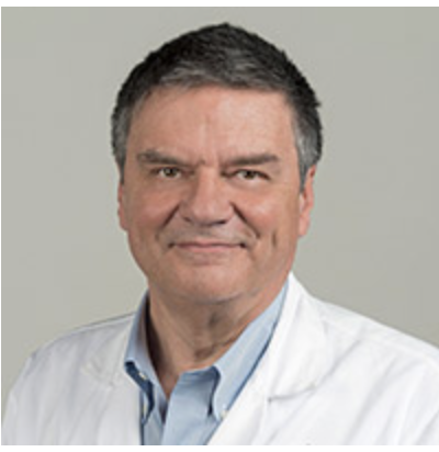
minutes, L and D. The speaker will discuss neuro oncology through interesting cases. The speaker is Dr. Moneer K. Faraj, President
0:47
of Arabic, Board of Neurosurgery in Iraq
0:51
Department of Neurosurgery, College of Medicine, University of Baghdad, Iraq. I will start the next session. talk from Dr. Munir Hamas Faraj, he's the president of Arabic chapter of the Arab
1:08
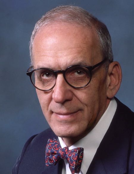
board of neurosurgery and he's eminent in neurosurgeon practicing in neuroscience teaching hospital back then Iraq. So welcome Dr. Munir and thank you very much. Okay, thank you very much for
1:23
inviting me, my dear professor, James Osman, my spiritual father, I've delighted to have my dear colleagues, especially Dr. Samar. When Dr. Samar invited me about this to talk about the
1:38
neuro-oncology, actually it's a very wide, huge subject and our aim is to focus on the young neurosurgeons and to make it an opportunity for more and make it more educational. Actually, I choose a
1:55
topic, a single topic and present several lectures about it. So I will talk only about the craniofarin geomas. I will give a brief fact and then I will describe four cases. The first case will be
2:12
with almost total removal and the second with a difficult removal. The third one will be very giant with partial remover and the fourth one was some of intraoperative complications.
2:26
Craniofarin geomas in general it's not so common. It's almost up to four percent of all brain tumors. Half of the cases will be injured and we have to buy more of the situation between 5 to 15 and
2:41
50 to 75 years. The symptoms are the onset either visual symptoms or those related to res intra cranial pressure and sometimes related to hypopituitarism. And children they may manifest as a delayed
2:58
a Pew Perti states due to a lowering of the growth hormone deficiency. And adults may be associated with decreased libido, amineurias, infertility, and the larger tumors where they extended to the
3:13
frontal lobe, they may have behavioral or personality changes. From the pathology point of view, it is regarded as a grade one, according to the WHO classification And most of the tumors have a
3:29
solid and cystic component and the cyst usually contains cholesterol crystals. Histopathology, it is either called adamame nematas, which is mostly in chilidin in this, the first slide,
3:48
and the oarpapillary, which is usually in the adults. And it's sometimes difficult to be differentiated from the
3:54
rascus cleftsis, only by immunohistochemistry.
4:19
techniques. Radiologically, usually, the cranial pharyngeomes have a heterogeneous signal zone, both T1 and T2. And it is more common than the pituitary that it has a supercellar extension. We
4:19
can differentiate it from the epidermoid by the fact that it will not have restricts restriction on diffusion, weighted image, and may be differentiated from the rascus cleft cyst by the fact that
4:33
this cyst is usually has a no solid mass and the enhancement will be limited only to its wall. Cranial pharyngeomes usually manage surgically still the golden standard for the cranial pharyngeomes is
4:50
the perional cranial tummy because this approach will let us able to see the more
4:58
important structure that we may face, including the optic nerves, the carotids, the laminator menalis, and even it will bring us a chance to pass it through certain corridors as I will show you in
5:12
videos. Usually, we aspirate the system debug the tumor and then we try to remove the solid part by a piecemeal and avoid retraction, especially to the posterior to the area of the hypothalamus,
5:28
and I usually call it the heart of the brain whenever you do over manipulation on the hypothalamus. Usually, the patient will never regain his consciousness after surgery.
5:42
Still, there's other approaches like sub-frontal. Sub-frontal usually, I do not like it. It has a limited space, and usually, it may be used with cases with a prefix
5:56
of the chiasm. Transcalusal, F. the tumor was exclusively within the third
6:01
ventricle. And those copy may be used to help us to visualize the lateral aspects of the operative field where the microscope cannot show them. And the transphenoidal approach, I tried it several
6:14
times. It may allow you for chiasmology compression, but still it has a very high risk of CSF leak post-operatively
6:24
Cranopharyngeum is actually has a role in radiotherapy. And focal radiation, whether after growth total resection or sub-total resection, have been approved to some sort of more control on the
6:41
tumor growth. Those with cystic tumor, several triers have been done with intracystic administration of
6:49
radioactive isotopes like euterium, but actually we never did that in our.
6:56
practice, stereotactic radio surgery and especially the gamma knife can be used only for the solid part of the mass and usually must be at least more than one millimeter away from the optic apparatus
7:14
and usually we have to expect there will be a certain reaction to this radio therapy with an increment in the size of the mass and the first five months following the radio therapy. In general, in
7:29
cranial pharyngeal mass, the best chance to cure or long-term control is your first surgery you did to the patient and the five-feet survival is arranged between 55 to
7:43
85 percent. During the description of the four cases, I use the Moeller-Vedel microscope. It is installed in our neuroscience hospital since
7:56
2002. The camera was a personi, but I changed it. It's not an HD I had to buy from Amazon. This is what we call lensless camera. Take the picture from the microscope and make it an HD picture and
8:14
send it to my laptop where I record the surgery and then also put it on a TV screen in the theatre so that my colleagues and the students can see what I'm doing and in order to make the record of
8:31
these operations and make them on touch, I had to learn myself the Adobe Premiere and the final cut-up role in order to make what I'm going to show you In the case one, I did it actually three weeks
8:49
ago. It is a four-year-old girl presented
8:55
with headache. vision, worsening, and other parts of its examination were normal. Her cranial
9:02
MRI shows this isrogynous mass. It does not reaching to the third ventricle, but occupy the whole cellular area.
9:15
We started with the right periodontal approach. And
9:19
I use what I say, I call it pull and leave, just to see and
9:26
to understand the attachment of the nature of the attachment of this tumor with the nearby structures.
9:35
Then I use
9:41
this microdisector, it's called a multi-disector, in order to separate the tumor from nearby tissues. Usually it is best to start to debug the tumor from the contralateral side. Here I open the
9:51
right periodontal approach and I start to super. the tumor from the left optic and carotid apparatus. While I'm pulling the tumor slowly, I'm trying to notice if there is any associated movement of
10:10
the nearby vessel and nerve so to avoid pulling them with me. That's why we call it in a very quiet piecemeal technique.
10:32
Then, we have the last part on the epsilon total and nearby the hypothalamus
10:41
It is almost completely removed.
10:48
Here, you should be very careful not to make any other pleasure on the hypothalamus.
10:56
And while pulling, you have to see if there is any abnormal movement with your movement for the results and the nearby nerves.
11:06
This is almost the last part
11:22
You can see the carotid and the
11:29
contralateral optic. Now it's almost removed. This is the optic of the epsilateral side.
11:36
Are the patient fortunately done well postoperatively? In the next case, we have a 17 years old male with headache and reduced visual equity As MRI showed, this mixed density mass occupying the
11:51
cell and reaching to the third ventricle.
11:56
With this all part, actually, at the bottom of this tumor.
12:03
We started also the right abdominal approach. This actually had the previous surgery. So we face adhesions, postoperative adhesions of the previous surgery, and I had to open the sylvian in order
12:18
to drain more CSF to - make the brain more relaxed. This is the olfactory group and we start now here to see the optics.
12:34
Now, we see both optic nerves and the optic chiasm is a prefix though we start to make an arachnoid dissection
12:46
and the pre-chasmatic space,
12:49
but nothing actually have been seen so clear and I created a bleeding.
13:05
Since I didn't show any significant tissue, I just handled with this bleeding, and then we went back to the laminar terminalis. We start to open the laminar terminalis area, and you can notice the
13:17
machine oil fluid start to come
13:40
Here, we feel there is a solid part, a huge part, that cannot be gated out either from the pre-cosmatic space or from the opening of the laminar terminals. I try to push it from here and there and
13:58
try to crush it, but I couldn't Then I use this ring curate to separate it more from the nearby structures, especially the carotids and the internal service of the optic chiasm.
14:17
Actually this is not an easy maneuver and it really needs a brave heart to do it
14:28
And then
14:31
we push from laminator menalis, push it more towards the pricky asthmatic space.
14:39
and we try to remove it through the prismatic space.
14:47
It's a very huge one
14:58
It takes your heart with you,
15:02
then the rest of the tumor mass, it's actually soft, have been removed through the laminator binalis.
15:15
Believe it or not, these videos made with and made
15:21
in China, camera, which costs only 100. Now
15:29
it comes clear.
15:32
We put surgery for hemostasis and this is the postoperative MRI with almost complete removal and the solid part here is just the piece of the surgery that we put in
15:48
The third case it said 12 years old male presented with head hypnosis and the blood vision was such a very huge.
15:60
mass extending towards the brain stem, even towards the lateral ventricle. I did just an coronofar angioma. We did right tiptineal approach. The coronetomy was a little bit difficult. That's why
16:13
we have a lot of bleeding. But just when we try to retract the brain, the tumor appeared immediately
16:26
There is the calcified part of it.
16:30
And a part of this is also appearing in the supercellular space. We try to open it.
16:39
And a very huge amount of this machine oil fluid came out.
16:47
It's very huge
16:53
We use two sectionitudes to aspirate it in order to prevent the spillage of this machine oil to other areas to create a chemical maningitis
17:08
The beautiful thing here, you can see that there is a calcified part coming out of both optic nerves in the pricky asthmatic space and there is another one between the contralateral optic and the
17:23
contralateral carotid.
17:26
So we try at the beginning to do dissection I usually start with the contralateral cells, so to dissect, remove, try to superior the tumor from the nearby carotid and the optic nerve, but actually
17:43
it was calcified and very hard,
17:48
then I try
17:51
to manipulate the other one
18:01
between the two carotid nerves and the prekey as much space Yes
18:10
but cannot be removed. So I use this biopsy for steps in order to crush the mass and try to remove it in piece mill. Believe me, these manipulations are not easy. How much you should
18:30
put off your load on the optic chasm and nerve.
18:39
I just have to get him out
18:45
We continue
18:54
And this is an epsilateral one
18:59
between the carotid and the optical carotid corridor. Also when we pull, we see the movement of the vessel. If it comes with it, we leave it. And this is the subtotal removal of the mass per
19:13
separator city. I couldn't remove the whole solid part because of its attachment with the carotid, especially on the ipsilateral side. The last case is a 20-years-old male presented to the blood
19:28
vision and behavioral changes. His MRI shows this huge mass of cuban referred ventricle and
19:37
acetortic care with mixed density. We did the surgery We started at the pre-cosmatic space,
19:48
but I saw this.
19:54
I saw this something. I don't know what it was at the beginning. It is just either a cystic maras or a vessel, but it's cross reaching to the area of the optic chiasm. You see this bulge. I mean
20:08
this bulge. So what is it? I couldn't know what is it actually at the beginning of the surgery. So I kept with my dissection of the arachnoid
20:23
Yeah.
20:26
Then
20:29
I assumed it is the
20:32
tumor mass.
20:35
And I tried to function it. So I used this This is Mike and I
20:45
and the disaster occurred. Now there is no montage, it is the natural speed.
20:52
I taught them to bring another section to you.
20:56
I tried to control it. It was in the carotid, actually.
21:04
So this is the real work
21:10
They are preparing the bipolar coterie for me and they are preparing the other section at you
21:28
There it is.
21:31
I do try to control it with the bipyrecoterity.
21:47
and it stopped. I think I was lucky at that time.
21:54
But this did not make, we didn't find the tumor till now, so we continue our search
22:02
on the laminatorbinalis. We opened the laminatorbinalis and the machine oil appeared. So actually I reviewed the journals later on and this case was a reopening for the cranial pharyngeal. It's not
22:19
the first surgery and I was able to have two papers saying that after you do operation for cranial pharyngeal or pituitaridinomas, you may have a fusiform aneurysm of the internal carotid, which I
22:38
face in this case. Now the calcified materials start to come out.
22:48
I try to remove as much as I could
23:12
Another assist
23:35
We find a chat it
24:02
And this is the first operative result, with also subtotal removal. And thank you very much for your listening.
24:13
I would like to congratulate Dr. Munir Farash, because for his honesty, outstanding, it is very unusual to have a neurosurgeon show a complication, it is very unusual for a neurosurgeon to show
24:30
that he left some tumor behind. We all go to different conferences and come back home wondering what I have done wrong. How can I take it all out the way that guy did, you know? I must do
24:46
something wrong, I must do something wrong So it is, I mean, I say that for the students, I mean, they think I'll stand that off.
24:59
Dr. Munir Farahrin sharing his thoughts and complications, and that is the wonderful thing. I think that, and independently, as I mean, to almost or not to almost, I mean, I guess that all the
25:10
consent was obtained from the parents. And anyway, the congratulations for the presentation where you show complications are not less than a perfect result. It's a perfect surgeon who can do that.
25:26
Congratulations Thank you.
25:30
Thank you, Professor Zaref.
25:34
Anyone has a comment or question now? I agree with Jorge. Dr. Manir, that's what Jorge said is exactly right. We had an SNI digital meeting about a year ago where there was a
25:54
Dr. Dio Pejari from India. who is showing
25:60
a transnasal endoscopic removal of the vaquenia fringioma. You mentioned that at the beginning and that it's, and you mentioned honestly that it's difficult to do and he showed a case of where he
26:14
was able to
26:18
get the tumor out. And then the problem is with, I consider this the most probably the most difficult tumor a neurosurgeon has to deal with. Because
26:30
the tumor is teasing you. It's teasing you because it looks like it's easy to do, so you go do some more, and you do some more, and then pretty soon you've gotten in trouble. And the
26:45
biggest question is, when do I stop? And you've covered all those things. And
26:52
so there are many different ways to look at this.
26:56
but he's looking at a transphonoidal. The beauty of that approach is you see everything from below and endoscopically, you're looking upright into the center of the tumor. The problem is, as you
27:11
mentioned, is how do you get all this to heal and not have a CSF leak as a complication which can be a disaster? So what's the right thing to do? Well, we need to have a case reports of a hundred
27:26
done with Dr. Munir's approach, maybe a hundred done with Dr. Monty's approach, maybe a hundred done with Dr. Patel's approach and a hundred done with the others. And not follow them just for a
27:39
day or a week, but follow them long-term and see what the consequences are. And long-term consequences of this disease are disastrous. And
27:49
there's a surgeon in New York who was very aggressive and wanted to take everything out at once So he gave the patients all the deficits immediately. Well, I'm not sure I want that.
28:02
And then the other approach is to come back and I had a patient where they did a right-tary anal quinionomy, then a left-tary quinionomy, and because you're forced to do that. And so what the right
28:14
answer is? I don't know what the right answer is.
28:18
But I commend you on doing that.
28:24
And you have to do what's best for you and what you can best do. And I agree with hurry. Everybody could show good results. What's the truth?
28:51
You need to be a very good surgeon to show you're less than perfect results and which were very good results in here. But yeah. Dr. Patel, can you try and talk about it? There's some meta,
28:54
there's some genetic differences. in the craniopharyngeal my maybe you can tell us about that yeah so I was going to say I commend you for having a very honest discussion about craniopharyngeal
29:10
hummus those cases are incredible I mean those are those are next level craniopharyngeal hummus really and and in neurosearch gone college in the last 10 years is from uncompunctional balance has
29:22
gained increasing usage and it is this idea that it's it's not just a perfect MRI we need to have the the patient at the forefront and so just because MRI looks amazing and all the time is gone if the
29:35
patient has DI and gains 200 pounds in a year and is blind you know that's that's a periodic victory and so it is important to understand particularly for craniopharyngeal hummus that are so terribly
29:49
disabling that 90 resection might be exactly what that patient needs you know relieve the mass effect, get them in a better state neurologically, treat the disease that's very adherent to the
30:05
hypothalamus or to the optic apparatus, either with breaking therapy or stereotactic radiosurgery or something else and keep the patient whole. You know, that is much more important than having a
30:18
perfect MRI and a hurt patient. And so I think, you know, cranio-fringiomas are a very good example of how to think about unco-functional balance in a real way. And then as Dr. Austin said, you
30:29
know, most cranio-fringiomas are at the adamantinomatous subtype, but there is a real percentage of cranio-fringiomas that are
30:38
the papillary subtype. And Priscilla-Brostianos out of Boston wrote about these a lot in the last 10 years in the papillary cranio-fringiomas. Their hallmark mutation is a BRAF V600E mutation. And
30:51
so for those patients, you can treat them with v-rathletes. inhibitors, MEK inhibitors, usually they treat them with dual therapy and the results are spectacular. Now, this is early data and who
31:01
knows what the long-term durability of that approach is, but they have, they had a series of case reports that came out of Boston eight years ago, maybe, of this terrible papillary cranial
31:14
friendeoma that every six months basically present into the ER, same patient trying to die because they're turmeric, they had a good resection, then their tumor grow back, then they're blind,
31:23
then they're hydrocephalic, and they're, it was just cyclical, and they're just beating themselves in the head trying to come up with the solution back to the OR, back to the OR, bilateral shots,
31:33
this, that, and the other, and everybody, I'm sure, has had a patient like that. And then they did next-generation sequencing, found this BRAF mutation, and treated them with the BRAF in
31:45
Meccan Hibber, and the tumor disappeared in six weeks, in six weeks, you know, something that, that had been life-threatening for this patient, times over over a couple year period. And then
31:55
they defined that many of these papillary cranial friendeomas have the same mutation. And so I think it's important as we think forward in neurosurgical oncology, if there's particular diseases that
32:07
we don't have perfect surgical answers for, maybe there's going to be something else, you know, and not to be blind about that.
32:15
Do you use the chemotherapy obviously after you've operated, but imagine the question that is when, when do you use it, do you use it after there's more recurrences, and because you're not sure
32:29
what the long-term outcomes are, what's, how do you approach that? I think, you know, papillary cranial pharyngeomas look different radiographically than adamantanometers, so you can know going
32:40
into surgery, and not perfectly, but with a fair degree of certainty, which of the two you're dealing with And so, for me,
32:51
if it's a papillary cranial pharyngeoma radiographically, and. I'm in the operating room and I think, listen, if I push this margin, it's really going to be detrimental to the patient's health.
32:59
Then I stop and I just would feel very comfortable with treating them with
33:05
medical therapy immediately after surgery, after they peeled up. If, you know, the surgery is going fabulously well, the tissue planes are easy, it's dissectable, it's not a particularly
33:15
technically challenging case, which is not often the case with cranios, but let's say that's the case, then I think you should go ahead and finish, you know, safe maximal resection, but, you
33:25
know, papillary cranios are the ones that are more solid in appearance on preoperative imaging. And so if you have that situation on MRI and you can define it for yourself, then I have no
33:37
hesitation about subtitling that subtype.
33:44
There was a report many years ago by a Dr. Shillitol. I'm sure Manir, you know this. He's worked in Boston with Dr. Mattson and he did histologic sections of cranial friend jomas into the,
34:00
obviously the patient's side, histologic sections into the hypothalamus. What he showed is that the tumors are very invasive, but some may not be, but you don't know, and in some cases you could
34:16
devastate the patient with what Dr. Patel was saying, the postoperative results are how do you, how can I explain to the family the patient's blind has diabetes and all kinds of endocrine
34:28
abnormalities and that's, I'm not sure the treatment's better than the disease there, but anyway, so I think your opening talk where you said that is a very key point. And a lot of people don't
34:44
understand that. It is, you think it's a circumscribed tumor, but it's not. And that's why I say it's, I think it's an extremely difficult team. Dr. Madi, your
35:05
thoughts?
35:10
That's a great job by Dr. Munir. Thanks for your marvelous lecture. Thank you very much.
35:20
Thank you. I think the
35:26
sequence of presentation of cases and the message behind the presentation that not every Cranio-Franjoma is a Cranio-Franjoma is obviously there for the young generation for those who are thinking,
35:41
trying to link the, let's say the biology, the histology, the pathology to the real surgery, I think Cranio-Franjoma is the shark of tank, even during residency if you want to, that you are
35:55
mastering or understanding the approaches. So Cranio-Franjoma with all the possible corridors, it's a surgical challenge. At the same time, everyone knows that it's stick to the neurovascular.
36:08
structure and it's not an active TMR. Even for oncologists, it's always a challenge. I think that's very a full discussion. Thank you for the expert opinion from the front part and thank you for
36:23
the opportunity to bring this point there.
36:28
We hope you enjoyed these presentations
36:33
Please fill out your evaluation of this video to receive CME credit.
36:43
The series is supported by the James I. and Carolyn R. Augsman Educational Foundation, owner of SI and
36:54
SI Digital and the Waymaster Corporation Producers of the Leading Gen Television Series, Silent Majority Speaks, Role Models, and Medical News Network.
37:12
The recorded session is available free on snidigitalorg.
37:20
Send your questions or comments to osmondsnidigitalorg.
37:32
Thank you


