0:01
Hello, I am Jim Osmond,
0:04
and welcoming you to another in the Sub-Saharan African Neurosurgery Grand Rounds series, sponsored by SI, Surgical Neurology International,
0:18
and SI Digital Innovations and Learning, a new video journal, interactive with discussion, in association with the Sub-Saharan African Neurosurgeons, Sub-Saharan African Neurosurgery International
0:36
Grand Rounds. This is the 10th meeting, held the first Sunday of each month,
0:44
and Matuso
0:47
is going to present a talk from the Kajabi Mission Hospital in Kenya on their experience in the treatment post-year foster tumors.
0:60
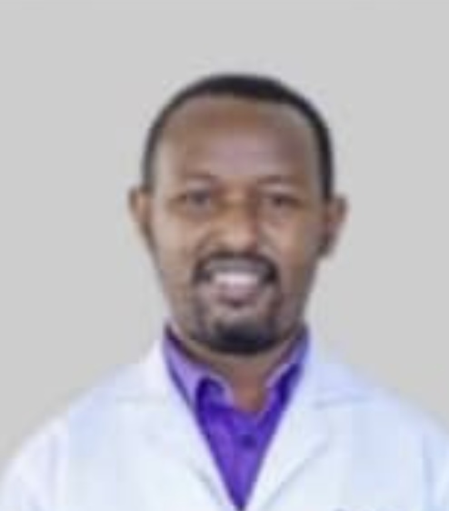
Ben is at the university hospital in Nairobi in the department of neurosurgery, and is at the Kajabi Mission Hospital in Kajabi, Kenya. So my name is Ben Jesso-Arman Yersajan from Kenya, and I'll
1:18
be presenting about our experience with posterior force achievements.
1:23
And this comes after a very high-powered presentation by Professor Elbaba, as opposed to his presentation, we'd not have a lot of technology, but we try with whatever facilities that we have Um.
1:44
So, I'll start a little bit on the epidemiology of post-reophosphatumas in Kenya, and like in many other parts of the world, the main age diagnosis is between 6 to 7 years, a study done here by Dr.
1:59
Peter and Jerry in 2020. And
2:01
so this is just a snippet on how we manage hydrocephalus in most of the children we see with post-reophosphatumas
2:08
Our first choice is placement of interpretive external ventricular drain that is gradually wind off in about 5 to 7 days.
2:19
And in most of these patients we are able to have them not be shunt dependent after surgery
2:31
However, this is not always the case as there are
2:35
many delays to definitive surgery and a lot of times is the patients have to wait for surgery. of resection of the posterior fossa tuma,
2:46
we prevent deterioration by placing a shunt fast as they wait for elective and surgery because of limited theater space. And therefore, we find ourselves at placing
3:00
ventricular peritoneal shunt preoperatively. And this allows us time to have, to sort of convert it from an emergency to an agent case.
3:12
And because of this practice in studies that have been done in our largest referral hospital, I came about 82 of children
3:24
had a VP shunt placed.
3:28
So that's a huge majority of the posterior force of tumor children ending up with ventricular peritoneal shunt There are centers that
3:40
practice routine routine
3:43
ETVs are before resection of posterior for sedumal, but that is not a practice that has picked up in our setup. So I'll present a few of the cases that we've done, that we've done here. And I'll
3:58
start with this case over 10-year-old Karl, who presented to us with a two-week history, who once did he get
4:05
an headache and vomiting We did a CT scan that showed a fourth ventricular tumor
4:14
that was hyperdense or non-contrast CT scan, and with upstream obstructive hydrocephalus with preventricular seepage. And we see the same on this sagittal cut of
4:31
MRI. So we placed a shunt before, because we could not go him gently and resected the tumor. And then after we took the patient, to Saje two or three weeks down the line. Interoperatively, we
4:47
found this tumor that was nochula
4:51
and sort of farm and we could get, we could go to a big chunk of it.
4:58
It's an envelope resection and at the end of the resection,
5:06
we could see
5:11
that the middle of the bellon, and beyond it, we can see the opening of the archidactors of
5:15
the cells and of CSF that was jetting from the aqueduct of cell vias. And really, we want, in this shows us that we've gotten the most cranial portion of
5:27
the tumor and this came back as a
5:36
metalloblastoma
5:39
the post-operative scan should. across the Tory section,
5:45
and obviously with resolution of the Hydrocephalus, though we still have our ventricle of Peltonia shunt in the larger ventricle. I'll go to
5:58
the next case of a 12 year old girl that I managed about two years ago. And she presented again with a four month history of gate instability, headache and vomiting We did a city scan on her and
6:15
can see that there's a large posterior for satuma
6:20
that is compressing this rebellion and displacing it
6:25
on either sides. And it seems to sort of split it in the middle line along the vamis and you only see strips of normal cerebellum on either side. Now this patient could not afford to do an MRI. So
6:37
we really just had to work with what you have and we have to walk with this city scan. of importance is that you'd see that this tumor goes beyond the cerebellum and seems to partially encase the
6:51
bacilla artery. And at this point, we could not really pick the distinction between the tumor and the brainstem. So we offered them surgery, but we were ready really for a subtotal resection
7:05
because from this the CT scan it appeared that there was invasion of the brainstem.
7:12
When we went for surgery, we found that there was actually a surgical plane between the tumor and
7:20
the pons. And the tumor had actually gone through the front end of Lushka into the purpontin space and of the batting on the bacilla artery. And through microscopic resection, we were able to reset
7:39
the whole tumor. The patient initially but you can see this is the post-operative scan. This is the Ali post-operative scan that we did on the first day, after surgery, which shows the cross-toto
7:52
resection. And this is a scan that we did on the patient about a year, about two months ago, which is nearly two years down the line, that shows cross-toto resection of the tumor and no recurrence.
8:06
And you see, and along with this area, you can see the widened from and of Lushka there, where the tumor had gone through into the prepointing space. So again,
8:22
quite an interesting case. And this is just another demonstration
8:30
of the extent of tumor before surgery and after surgery. Then what was the histology of that case? And this actually came back as a pile of citicastrocytoma.
8:44
really not the typical presentation. I was also lead and the pattern of spread was also initially with you. Over the boy's symptoms,
8:57
how did he present and how is he doing now two years later? Initially he had presented with
9:09
an ability to walk and actually when
9:14
the child was bound on a wheelchair, was vomiting and severely wasted and probably from the protracted period of vomiting
9:26
and also had inability to swallow. I would call it a difficulty swallow and had actually been treated severely for aspiration and pneumonia from the brainstem
9:43
After surgery, we had
9:46
to put a
9:49
gastros tummy for this patient. And she did well, although she had to keep the gastros tummy tube for almost two months after surgery. And slowly, she was able to get back her swallowing reflex.
10:04
And she kindly is able to swallow and has gone back to school.
10:11
Yeah.
10:13
Excellent. And I assume you did a midline approach or was that I can't see from the pictures here. Yeah. Yeah. Yeah. I'm sorry. I didn't bring any interpretive. Okay. It's okay. It's okay.
10:31
It's okay. Not to me. So very good result. It was terrific. But it took time. And I assume it took your time I just from the delay between thinking about an HEV and putting in a shunt, there
10:46
must have been enough some days before the patient was able to go to surgery. Am I right or did you ever able to go right away?
10:56
No, we had to take a bit of time and actually when the patient came to us, they had an active lower respiratory tract infection from aspiration because of the difficulty in swallowing So initially
11:12
we just put in a shunt and had to wait for the patient to be stabilized for about two or three weeks. Did the patient neurologically get better with the shunt?
11:24
The swallowing did not really get better. It was only on fluids and that time we put an
11:32
NG tube. Yeah, so the neurologic deficits did not improve with the VP shunt. The one thing that went away was the vomiting was also one that I quite a bit. Oh, another case similar to the one
11:47
that Sam are presented of extensive brain stem compression that recovered, it's just terrific.
11:56
Yeah, thank you. Yeah, and the last case is a four year old boy
12:04
that we managed about four months ago.
12:08
And this young boy came with an ability to walk for the last three months, had all the milestones that had progressed, was unable to talk, unable to sit.
12:23
And when they came, they were bound on the wheelchair again. So
12:29
we did a scan on them, and this time we could do an MRI that showed a large posterior forcetumor,
12:37
which appeared to be mid-line, solid and cystic components. And if you look keenly on the T2 axial cuts, you see that this compression of the fourth ventricle. So this really appears to be a
12:50
Vamiantuma
12:52
that is pressing on the fourth ventricle and the supertentorial obstructive irisyphylus with a lot of very ventricular seepage again.
13:03
So this patient, and on the
13:07
sagittal cuts, again you see the large posterior fossa tuma with a
13:15
solid and cystic complement that is pressing on the fourth ventricle and the obstructive irisyphylus. We were able to take them sagittal in good time and we avoided placing Vamiantuma on this
13:30
particular patient. And in Vamiantuma sagittal, we put an PPD
13:35
at the Dundee's point And
13:38
this enabled us to relax.
13:43
the contents of the posterior fossa, because the posterior fossa was very tight.
13:49
And what we initially thought that was actually cystic, a tunnel that most of us also saw it, but there was a good brain tumor interface and we were able to excise
14:01
the tumor. And this is a CT scan that we did on this on the first day of the surgery. And you see the fourth ventricle now is well visible. And that's the resection cavity showing again across
14:14
total resections. At this point, we still had our external ventricular drain in place, but over time, we were able to win it off and avoid an avoid a ventricle, a V-patient in this patient. So
14:29
again, just a comparison of the preoperative and the postoperative scan. And I have a feeling that our tumors are probably, I don't know what you see there, but we feel that all. mother probably
14:42
larger because of the longer duration it takes for patients to get actual care. You're right. What was the pathology here? The pathology again was
14:57
a pylocytic castrocytone. Okay, another pylocytic castrocytone. How did you know, as you were approaching it through the tumor, how did you know where the force ventricle was?
15:07
So from the
15:09
from the preparative scans, I had anticipated that it was anterior to the tumor, because as you can see on this G2 cut here, you can see, if you can see my pointer, that's where the photoventric
15:24
goal was. And I knew that I would,
15:29
it would be the last thing I would see. And once I saw the Tila Corolla on top of that forms the roof of the photoventric goal, then I knew
15:45
I'm assuming you didn't have any electrical recording or wasn't available, is that correct? No, we did not have any interpretive neural monitoring.
15:57
Yeah, but it's something that was readily available. We would have liked to use. Did you see the when you opened them up? Did you see the foramen magnum? Were you able to? Did you go that low or
16:07
not?
16:10
Did you go that low or not? Yes, when we did the suboxipital craniotomy, we did a large suboxipital craniotomy. And
16:19
you can see my bonicat
16:22
from the point from where the confluence of sinuses would be
16:29
all the way to the posterior remove the foramen magnum So, we removed quite a sizable portion
16:38
of bone and. And this was really to create
16:44
space so that we are not working in a tight space because we anticipated that this was a big leash.
16:51
I don't know if you use this or not. One of the tricks I used and maybe Nim knows about this, is you take a cottonoid and usually it has a black strip, you know, a black marker right on the center
17:01
of it. And you slide that in between the tonsils on the bottom and you slide it up into the fourth ventricle.
17:12
And if you can slide it in there, what happens is you're approaching the tumor from above. What you do is you wind up seeing the cottonoid before you get to the brainstem.
17:23
You understand what I'm saying? Yeah, yeah. So it's a way of keeping you away from the brainstem. If you're not sure where it is and use a cottonoid in there and it can help you. I don't know if
17:35
that would help be helpful to people or not We did that, I think it was helpful. So I think that was a terrific job.
17:44
Yeah, yeah, thank you.
17:48
How did the patient do? Yeah, so the patient did well, the patient
17:56
at least is able to sit now. They're still not working yet, but a lot of improvement in terms of the feeding and the motor function and he's starting to bubble, but the words that they could talk
18:09
before, you know, there was a global regression of all the milestones. So a lot of improvement from the last time that I saw the child. And how many months in surgery is it?
18:23
The child was four years old. And now, oh, four years old when he operated, how was he, you know,
18:31
how old is he now? Now he's probably, this was three months ago
18:38
Yeah, so maybe for. going to four and a half. So it's about six months, both surgery, is that right? Yeah. And you're expecting some more recovery, Sammer. I'm sure you've seen extensive
18:53
things like this. Do you have to sometimes wait a year or more to get recovery, full recovery?
19:01
Maybe Sammer's not here. What is trying
19:07
to do you have any answer to that?
19:11
I'm sorry, you're asking about the, sure, well, sometimes they can't take six or 12 months after
19:20
posterior fossa cases with impairment of the cranial nerve. No, is she being taken care of at home or is she in a hospital a bit?
19:31
Oh no, so
19:35
he is not cutie. He's, you know, he is not cutie issues that need to be done. that he needs hospital careful.
19:44
So there's a question in the chat been regarding comments on routine post-obscans and pediatric patients.
19:52
Is that done routinely with limited resources? Can that be problematic? What might be your frequency of follow-up scans after surgical resection?
20:08
Yeah, so
20:11
we do them immediately after surgery so that's only within 24
20:35
hours of surgery. And thereafter we are able to, we do them maybe every six months for the first three, three years, maybe a year thereafter, and annually thereafter for up to five years. In
20:35
terms of availability of imaging is a challenge sometimes, and you can see sometimes we have to cooperate just with CT scans.
20:44
And sometimes when you want to do a
20:48
post-operative scan, most of the barriers would be financial. So sometimes you want to scan, but the patients can't afford. But the imaging is available in most of the major cities, but not all
21:03
over the country Dr. Risterrata, can I ask you a question, please? Please. Either Dr. Ben, or you guys take the Baba, is there? Because when they do the post-operative surgery, sometimes
21:12
mutism happens for some time after the surgery. In this case, maybe mutism was somewhat involved in his recovery, or any comments some of the colleagues can
21:28
make. I really appreciate
21:32
that So, Sam, are you there? You want to make comments on a cerebellar mutism?
21:39
I think the, can you hear me? Yes. Yeah. I think the concept of, or the issue of staebella mutism or a better word to say this berserphos syndrome is now we understand it better than we used to.
21:56
And the old days we used to think that the surgical approach is the main issue. Cut vermis, not cut vermis. I think now we have better understanding that even the molecular genetics of the tumor is
22:07
now impacting the chance of having Pussyafossa syndrome or not, specifically in middle of last summer brain tumors. We now know very well that a higher malignant molecular profile tumors like group
22:22
three or group four can have a higher chance of Pussyafossa syndrome or cerebellar mutism compared to sonic hedgehog or WNT. I think - You have physiology behind it.
22:37
Nobody knows, nobody understands. I do believe those patients, but at the time they're diagnosed, if they're very malignant, they can cause venous congestion or some form of venous outflow
22:52
obstruction of the vermis and some of these bell or white matter tracts. Nobody understands 100, at least to my knowledge, I don't know. Has anybody done fiber track scans on these people? Yeah,
23:05
I've never done it for clinical reasons, but I know there's extensive literature out there about association of Pussyov-Hassa syndrome or mutism with specific surgical approaches as well as specific
23:18
molecular profiles of Pussyov-Hassa tumors. I personally,
23:24
to avoid Pussyov-Hassa syndrome, I focus on clean approach, maintaining venous outflow, Maintaining excellent vascular supply of venous alpha.
23:36
you're in the case avoid retraction and if you do have a long case to do intermittent release of any retraction avoid retractors as much as possible as well as avoid any vermion injury or vermion
23:51
splitting. We know that there's connections between the cerebellum and speech and that's why I asked you that and the fiber triage is not for balance. It also influences speech. Isn't that right?
24:08
Correct.
24:10
So, Samra, with your approach, have you found that in your experience that has made a difference with cerebellum, and as a corollary to that, if we'll posteriorly pause the syndrome, in cases
24:29
that you have had, if you've had any, were there common features,
24:37
Thank you, observed. It's a double itch question, Estrada. I had a, there's no question my practice over the last 10, 15 years, I have seen less and less cerebellumutism because I'm becoming a
24:49
more experienced surgeon. And I'm doing a better job of doing list retraction, quicker surgery, respecting the venous outflow and avoiding any significant cerebellar edema intra-operatively. At
24:58
the same time, over the last couple of
25:02
years, I
25:08
have had a wonderful straightforward, clean-cut edema and
25:15
non-existing edema cases. And the cases went extremely well. Post-up day one, the child is fine. Post-up day two, they start struggling with talking on by day three or four. They have full-blown
25:31
pusserphus syndrome, although I've done a wonderful operation. So I do believe we as, neurosurgeons, we don't understand very well how this works. At the end of the day, we have surgeons who
25:42
should maintain
25:47
an operation that achieves the goals while minimizing great traction and respecting the venous outflow. But at times, I can tell you, I've done a wonderful operation and the child's struggle for
25:59
six weeks with Pusserfossa syndrome. I don't understand it. All right, so there's some questions How is an endoscope has helped you with less traction, less trauma to the cerebellum? Great
26:14
question, Strata. The techniques I showed earlier are for non-Pusserfossa, classic mid-line Pusserfossa tumors. Yes, I put
26:23
the endoscope at the end of surgery, especially if the tumor goes, some appendamomence that go for immunofluidical or go too laterally as I put the endoscope
26:30
to
26:32
take a look, but it will not impact, in my opinion, to self-assist syndrome.
26:37
I think there's some questions. Is Strata for Ben at the end here? One is what approached you recommend Tila Vilar versus Transvermium and posterior fossa tumors?
26:50
Would you having a strict midline lesion make a difference in choice?
26:55
Maybe Ben and Strata could answer that
27:04
I
27:06
prefer to use the Tila Vilar approach for both ventricular tumors and like Professor Elba Baba has already alluded to, this really keeps the structures from any
27:23
damage during surgery. And therefore this seems to have, I don't know the argument about it, but this has a lower risk of posterior fossa syndrome
27:39
But when it comes to a case like this that we are showing now, which is a cerebellar, I think the approach is obviously not really
27:54
transcortical because the tumour is really showing on the surface of the cerebellum, but I think the way to avoid complications is to avoid retraction on this cerebella hemispheres.
28:08
Yeah, that's what I'd say. So now are any comments about the trans. the trans-helovular and trans-fermion?
28:19
I guess, I don't know if he's still here.
28:25
Okay, that's it. I wanted to know about how common do you see seizures and posterior fascia tumors?
28:33
I have not, I have, I have not seen seizures, seizures
28:40
in my, not seen seizures from, from posterior forces, SAGI, unless maybe they supertentoria spread over disease That's what I would think when I see that, but that's something that I have not
28:53
really seen in the absence of supra-tentoria pathology.
28:60
Let's ask a question to Professor Elbaba and thank, I would like to thank him for, I hope he's still on the board. Are you still with us? He may have. I still, yeah, it's a very good
29:15
presentation to give, but there's one comment he made. that is the endoscopic assisted is mainly for the largely placed tumors of the posterior fossa. So, and the mid-length, classic mid-length
29:29
tumors of the posterior fossa like the one Dr. Ben Moutis was presented. He does not, well, he seems to not recommend endoscopic assisted surgery for that. Did I get him clear on that? Because I
29:48
just like that clarification. And of course, that most of the cases he presented were classically or laterally blessed tumors, not the classic mid-length tumors, which Dr. Ben Moutisso has
30:02
presented. Then the other one, which the other comment, which I wanted to make is a very good presentations, one from a very, very well established unit in North America and the other one from.
30:17
a sub-Saharan African country, which is really trying its best to create good neurosurgical practice. And the, what is very clear is that we are still pursuing neurosurgery in the old fashion mode,
30:35
but still getting very good results. And what Professor Elbaba has presented, it will probably, I know that in some of our teaching hospitals, it can be attempted. And I know about two or three
30:51
teaching hospitals where it could be, that could be applied. And this is the other question I'd like to ask Dr. Ben Mutiso. Ben Mutiso works in, I think, two main hospitals, or maybe three,
31:07
which are public hospitals. And I think he does a lot of endoscopic work So, I would like him to comment. on the presentations of Professor Elbaaba, as far as his technical development is
31:24
concerned. Does he feel that some of these methods, which have been mentioned by Professor Elbaaba, since he's endoscopically very competent, does he see any pathway in which maybe he may apply
31:38
some of these methods in his future practice, Dr. Mutis? So the first question was for Professor Elbaaba Thank you for that wonderful presentation. But what I wanted to know is I think you say
31:49
that endoscopically assisted was mainly for the laterally press was therefore the tumors, but not for this classic midline, the tumors. Yeah, Professor, I do not see a - Roberta, can we stop
32:02
sharing screen here? 'Cause we're not seeing, we're just,
32:06
Ben, that's it. Okay, go ahead. Professor Nemiro, thank you for the comment. I do believe there is no significant value for using.
32:17
the endoscope for a pure medline, lesion, resected microsurgically. Yes, you can put the endoscope to take a look at the confirmed gross total excision, but I found more value in the pizia fossa
32:33
via retrosigmoid approach or via telovila approach for a tumor that extends laterally like an epindimomence to fereminoflujga and goes into
32:47
the lateral to the brainstem.
32:51
But I really want to make a comment for Professor Ben Matisse's lecture. First, I would like to congratulate him and his team for doing this excellent work with the resources you have to achieve,
33:06
clean, beautiful resections, keeping the children safe, doing everything in your power to reduce the morbidity and mortality. with those children with complex lesions. So I have nothing but
33:19
utmost respect and appreciation for you sharing this wonderful talk with us. Thank you.
33:27
Yes, I'd like to add to that Ben. That was a tour de force operating on that on that large posterior fossil tumor, just equipped with the
33:40
CT images. I mean, we're so used to being able to have much more definition with MR imaging, but you did a phenomenal job solely based on your, the imaging that you have pre-operatively just being
33:54
a CT scan. So congratulations, Ben. I agree. Anybody else have any questions? Professor, Professor J. Maan, or
34:04
anybody else there? Okay, I'd have a lot to ask Do you value CT angiography or MRN? in your 100 of these cases.
34:19
For your endoscopy. Do you insist for the pineal regentimals? Do you insist on an geography? No, I do not see a value for angiography except for selected cases where they heavily vascularized
34:38
lesions like haemangi blastomas, potentially some
34:44
interventricular, large choroid plexus papillomas or choroid plexus carcinomas. There are some people who do selective embolizations prior to surgery. I personally do not see a value. I believe
34:58
that surgical anatomy and a good technique is the way to go with whatever armamentarium of equipment you have in your hospital or operating room. Thank you, Dr. Mutee So Professor Nimrod has posed
35:13
a question to you regarding - your thoughts about the application of the endoscope with posterior fossa tumors as Dr. Elbaba has presented. What were your thoughts about the implementation of that
35:30
where you are in practicality?
35:36
Thank you very much, Professor Nimrod I think we can employ
35:44
a lot of the techniques that we have seen,
35:49
Professor Elbaba show us. And in a few of the facilities that we have, we have flexible and rigid endoscopes that we use for ETV and CPC. So this, but what we lack are just a few things that we
36:10
can add onto, like endoscopic equipment, like microscissors.
36:16
and the bovis and the likes,
36:19
but seeing that we are able to do hit TVs and CPCs and we are able to do biopsy for binary endoscopic biopsies for pineal region tumors, I think this is a practice we can push a notch higher and take
36:35
it to the level of professor at Baba and maybe with his support and mentorship, we could be able to take the endoscopic quack to the next level,
36:48
thank you. Well, Sam or we might have to arrange for you to go out to Kenya to
36:55
lend a hand and help them get moving to the next level. I'm committed to help and partner with you all in any way, shape or form, thank you. Thank you, excellent, excellent. Dr. Sareta, I
37:07
have a question if you don't mind Dr. Elba or the other colleagues regarding the mutism.
37:15
well on medicine. There was a discussion that
37:18
perforators may be affected and that is the cause. Any comment on that?
37:26
What was, I didn't hear that aside, what might be affected? There's the perforators to the brainstems, maybe the cause of the diabetes, and that discussion was there, and I need some comment if
37:38
you can, but thank you If I may answer, to my knowledge, out of all the reading I've done,
37:48
and out of frustration, personally, in some cases, where they developed Pusserfassa syndrome after a clean, beautiful operation, if you look at the diffusion imaging on the MRI, you find it's
38:04
normal in most cases, which makes most people believe that more likely than not, it's Venus outflow congestion or or obstruction or some form of venous infarction that's not showing on the
38:23
diffusion imaging
38:30
that's contributing the edema from the retraction. Plus, as we discussed earlier, the molecular profile of this malignant tumour. We see no question. We see more Priscilla fossa syndrome with
38:39
malignant than benign tumours in the Priscilla fossa. So is there a component of maybe micro infiltration of malignant cells into the veins or causing venous blockage? Nobody knows. But I do
38:54
believe there's the pediatric neuro oncology groups are heavily invested in studying this phenomena. I know a few months ago, I saw there's a conference they they put together, which is a respected
39:08
initiative to discuss Priscilla Farsa syndrome. conference put together by, by neuro oncologists and neurologists and neurosurgeons. There's more and more interest in understanding it. Thank you.
39:23
Thank you. Thank you. Thank you, sir. No other comments. One other question. I think there's one in the chat. On the chat for Ben about most of
39:37
your foster craniotomy or craniectomy. What's your experience?
39:44
I have hardly done posterior foster
39:48
craniectomies and most times I have done craniotomies in the posterior foster and I always endeavor to put back the bone flap
39:58
and this really is because one is that you know you maintain that that plane and in the event that you have to go back then you are gonna have a good plane and you will not have the dura stack on this
40:11
aboxipital muscles.
40:15
And secondly, also, I think if you leave the post - I think if you do a cronyectomy, there is a high up -
40:35
I don't know what the experience of the rest of the team is, I know, um, um, um, um, um, um, my teacher, Professor Nimrod might have, uh, a mochum. I don't know. It's, uh, I think I'd
40:40
like to get for thousands And, uh,
40:50
I don't know. It's, uh, I think I'd like to get for thousands. And, uh, Professor Bernard's views on this. In the early hours, we used to be professor for the cronyectomy. And then that's
41:04
wrong. And then that's wrong. And then that's wrong. Of course, there was a cronyotomy was introduced. And, uh,
41:11
I think. Maybe my colleagues in the crime comment on that I mean, most of the patients who we did was here for a cleaningectomy. We never had problems. And, uh, when now. was elevated to
41:25
posterior fossa craniotomy. It was embraced in equal measure and the older neurosajuns were very resistant to moving to posterior fossa craniotomy.
41:43
But my colleagues, my other senior colleagues may comment, what are their views on this?
41:48
I think that's an interesting question. I can tell you that earlier in my career, and I had an interest in vestibular schwannomas, and I did retromastar
41:58
craniectomies for vestibular schwannomas, because that's how I was trained. But I must say I had a vexing problem that was with post-operative headaches in patients that I thought, well, but when
42:16
I converted to doing retromastar craniotomies, that headache issue resolved. I mean, it wasn't percentage but it was enough to be quite a frustrating problem for me
42:31
okay Jim any comment I know I see Alvin Albany is still with us Alvin
42:42
Alvin Doe
42:45
I'm yeah I'm following okay Albany have any comments about today I always look forward to your opinions
42:54
not really I think the presentations do were great it is just professor Ben Matuso regarding
43:03
the first case
43:06
they post-operative brain CT scan I noted that there were still a shunt so I just wanted to get a clarity whether it was to the EVD or the EVD was converted to VP shot.
43:24
Yeah, thank you for my question. For the fast case, it was a metal oplastoma. Then like I mentioned, when we are not able to operate them as soon as we want to, we would put in shuns just to buy
43:36
some more time. And that's what happened for that patient. So we put in a shunt and then we had to wait for two or three weeks before
43:45
we could get a slot to do operative resection of the tumor. So yes, that was a VP shunt and the patient still has to be patient as to whether they really need it. I don't, I think they could have
43:57
done without it. Yeah.
44:02
Samra, did you want to comment on craniectomy versus craniotomy? I think craniectomy
44:09
is only reserved in patients with
44:14
unusual intraoperative severe cerebellar edema that you don't feel comfortable closing the door out
44:24
I almost never do craniectomy. Now, craniotomy is the way to go. Now,
44:32
for colleagues who continue to do craniectomy, it is okay because the suboxymptom muscles will always provide good support and protection. The issue is that if you have a wide vasophosa craniectomy
44:49
you can end up with the sagging where the
44:58
suboxymatic would be sagging extra crannulae. And I had to do one case a few years ago, a patient who had a wide Chiari malformation, Chiari craniectomy where developed lower crannion or apathy is
45:15
from the sagging and ended up doing some cranioplasty to help with the symptoms, which was successful. So I do believe if this surgeon has the ability to put the bone back, do a craniotomy rather
45:29
than craniectomy, and secure it with whatever resources you have, sutures or micro plates. I do recommend it. It's better long term. And also, if you come back to take care of a recurrence after
45:43
two, three or five years, there's a tumor recurrence, having the bone will create an easier approach, because the suboxptom muscles can really scar down into the tura, and can be difficult when
45:56
you go back.
45:59
Yeah, I see. I
46:02
just rather it's 10 o'clock. We've gone in for a couple hours and terrific discussion. This is Sam, or thank you and Ben terrific job with very challenging cases and anything else you want to say
46:16
is try to know. I think this is an excellent discussion and thank both of you very much. Thank you very very productive very thoughtful. Obviously, there was very active participation reflecting
46:28
the quality. So thank, thank both of you very, very much. This was both of you to all of you. Thank you. Okay. Thank you. Well, thank you. We'll, we'll wind down and we'll, we'll be back
46:42
the first Sunday next month. Take care. We'll keep it going. Thank you. Thank you. All right
46:51
We hope you enjoyed this presentation.
46:55
The views and opinions expressed in this program are those of the author and interviewee and do not necessarily reflect the official policy or position of SNI digital arts management. The information
47:10
contained in this program should not be considered to be medical advice. Patients should consult their own physicians for that particular advice in regard to their or their. family-specific medical
47:26
needs.
47:29
Please fill out your evaluation of this video, which is located at the bottom of the video on the home page, on the rating scale as indicated below, choosing one to five stars, five the highest to
47:44
help us improve our program and the information we bring to you in SI Digital The foundation supporting these journals believes there are many ways to learn. SI, or Surgical Neurology International,
47:50
is an Internet Journal founded 15 years ago. Nancy Epstein, as its editor-in-chief, is what addresses SIglobal. And SI Digital is a new video journal of neurosurgery. It's interactive with
48:13
discussion exploring innovations and learning.
48:25
And its one address is SNIDigitalorg. Both are
48:30
free 247, 365 on the internet.
48:37
Surgical neurology international has been published for 15 years and has read in 239 countries and territories. And it's the third largest readership of neurosurgical journals in the world
48:52
SI digital has published basically for more than 12 months now. It's seen in 149 countries. And it's the first video journal in our surgery.
49:15
SNI, or Surgical Neurology International, has been published for 15 years and has read in 239 countries and territories and has the third largest readership of neurosurgical journals.
49:32
SNI digital innovations in learning is seen in 149 countries in 12 months since its publication and is the first video journal of neurosurgery. Foundation supporting these journals believes in
49:50
helping people throughout the world. This recorded session is available free on SNIdigitalorg.
49:59
You can click on the icon at the bottom right of the home page if you have questions, comments, or requests for CME
50:10
SNI Digital also is now offering all of its programs on podcasts. On Apple, Amazon, or Spotify, look for SNI Digital. The foundation supporting these programs also is a medical news network,
50:26
bringing truthful medical and science news to the world.
50:31
The programs are copyrighted by the James I. and Carolyn Oesman Educational Foundation, all rights are reserved. Contact James Oesman at the email address listed on the slide. We hope you enjoyed
50:45
the program. Thank you very much for watching.